Highlight
• Elevated plasma PGPC correlates with coronary artery disease (CAD) severity and poor collateral vessel formation.
• PGPC inhibits endothelial cell migration and tube formation by impairing autophagosomal-lysosomal fusion.
• NPC1 upregulation via CD36 mediates syntaxin 17 acetylation and ubiquitination, leading to its degradation.
• Targeting PGPC, NPC1, or syntaxin 17 rescues impaired angiogenesis, suggesting novel therapeutic approaches for CAD.
Study Background
Coronary artery disease (CAD) remains a leading cause of morbidity and mortality worldwide, often resulting from atherosclerotic plaque formation that limits myocardial blood flow, causing ischemic injury. Angiogenesis, the formation of new blood vessels from existing vasculature, is critical for restoring blood supply to ischemic tissues and facilitates myocardial repair. However, in CAD patients, impaired angiogenesis limits tissue recovery and worsens clinical outcomes.
Autophagy—a cellular process involving degradation and recycling of cytoplasmic components via autophagosomal-lysosomal fusion—has emerged as an important modulator of endothelial cell function and angiogenesis. The oxidized phospholipid PGPC (1-palmitoyl-2-glutaryl-sn-glycero-3-phosphocholine) accumulates in atherosclerotic lesions and has been implicated in vascular dysfunction. Yet, the impact of PGPC on autophagy-regulated angiogenesis, particularly in ischemic heart disease, has remained unclear.
Study Design
This study investigated whether PGPC modulates angiogenesis through autophagy regulation using a combination of clinical sample analysis, in vitro endothelial cell assays, and in vivo animal models. Plasma PGPC concentrations were measured in CAD patients and correlated with disease severity and coronary collateral formation.
Functionally, PGPC effects on endothelial migration and tubulogenesis were assessed in human endothelial cells. Autophagic flux and key autophagy-angiogenesis-related proteins were examined. In vivo, angiogenic capacity was evaluated using subcutaneous Matrigel plug assays, hindlimb ischemia models, and myocardial infarction models in C57BL/6J mice with and without PGPC exposure.
The interplay between molecules involved in autophagosomal-lysosomal fusion—specifically syntaxin 17 (STX17), NPC intracellular cholesterol transporter 1 (NPC1), and upstream regulators like CD36—was studied to elucidate the mechanisms by which PGPC impairs endothelial autophagy and angiogenesis.
Key Findings
Increased PGPC Levels Correlate with CAD Severity and Impaired Collaterals:
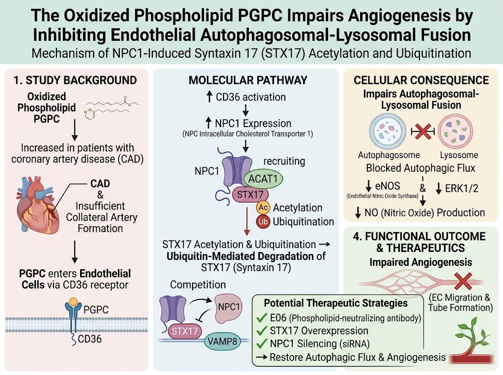
Patients with CAD demonstrated elevated plasma PGPC, which positively correlated with angiographic measures of CAD severity and negatively correlated with the formation of coronary collateral arteries that normally improve tissue perfusion during occlusion.
PGPC Impairs Angiogenesis In Vitro and In Vivo:
Exposure of cultured endothelial cells to PGPC markedly inhibited cell migration and tube formation, essential steps in angiogenesis. Consistently, PGPC treatment reduced neovascularization in subcutaneous Matrigel plugs and decreased blood flow recovery in mouse hindlimb ischemia models. In myocardial infarction models, PGPC suppressed angiogenesis in ischemic myocardium and significantly inhibited VEGFA-induced proangiogenic effects, highlighting its detrimental influence in ischemic heart repair.
Mechanistic Insights—Disruption of Autophagosomal-Lysosomal Fusion via STX17 Degradation:
PGPC suppressed the expression of syntaxin 17 (STX17), a SNARE protein critical for autophagosome-lysosome fusion, leading to impaired autophagic flux in endothelial cells. PGPC induced STX17 acetylation, which enhanced its ubiquitination and proteasomal degradation. This effect was mediated through upregulation of NPC1, a cholesterol transporter also involved in endosomal trafficking signatures.
Role of NPC1 and CD36 in STX17 Modification:
PGPC upregulated NPC1 expression via the scavenger receptor CD36. NPC1 recruited acetyltransferase ACAT1 to promote STX17 acetylation, facilitating its ubiquitination. In addition, NPC1 competed with STX17 for binding to VAMP8, another SNARE protein, further impairing autophagosome-lysosome fusion. This molecular disruption inhibited downstream signaling pathways including eNOS, ERK1/2 phosphorylation, and nitric oxide production, all critical to endothelial angiogenic function.
Reversibility by Targeting PGPC, NPC1 or STX17:
Neutralizing oxidized phospholipids with the antibody E06, overexpressing STX17, or silencing NPC1 restored autophagic flux and rescued impaired angiogenesis in endothelial cells and ischemic tissues. These findings provide compelling evidence for the causal role of PGPC-NPC1-STX17 axis in regulating endothelial autophagy and angiogenesis under ischemic conditions.
Expert Commentary
This study addresses an important gap by linking oxidized phospholipids commonly found in atherosclerotic lesions to the modulation of endothelial autophagy and angiogenesis. It highlights syntaxin 17 as a novel molecular target influenced by posttranslational modifications that govern autophagic flux and vascular regeneration.
The demonstration that NPC1 mediates a critical regulatory role in syntaxin 17 degradation via acetylation and ubiquitination adds a nuanced understanding of intracellular trafficking pathways impacting vascular biology. The involvement of CD36 linking extracellular oxidized lipids to intracellular signaling cascades underscores the pathophysiological relevance in atherosclerosis.
Although the preclinical models convincingly establish causality, further translational and clinical studies are warranted to evaluate potential therapeutic interventions targeting PGPC, NPC1, or syntaxin 17 in CAD patients. Consideration of possible off-target effects or systemic consequences of modulating autophagy is essential.
Conclusion
PGPC, an oxidized phospholipid abundant in atherosclerotic lesions, impairs angiogenesis by inhibiting autophagosomal-lysosomal fusion in endothelial cells, principally via inducing acetylation and ubiquitination-mediated degradation of syntaxin 17. This process is critically mediated through NPC1 upregulation driven by CD36. Therapeutic strategies that neutralize PGPC or restore syntaxin 17 function may promote angiogenesis and improve recovery in patients with CAD, representing a promising direction for future cardiovascular regenerative medicine.
Funding and Clinical Trials
The referenced study was supported primarily by relevant cardiovascular research grants. No specific clinical trial registration was provided for this basic and preclinical research.
References
Li L, Gao JJ, Kang YT, Pan WK, Jian YP, Liu YJ, Deng MJ, Cao Y, Lyu CY, Wu FY, Chen L, Liu JH, Dai G, Liao JB, Xie HJ, Bai YP, Zhou JG, Li Y, Ou ZJ, Ou JS. The Oxidized Phospholipid PGPC Impairs Angiogenesis by Inhibiting Endothelial Autophagosomal-Lysosomal Fusion Through NPC1-Induced Syntaxin 17 Acetylation and Ubiquitination. Circulation. 2026 Oct 6; PMID: 42836285.
Additional relevant literature on autophagy and angiogenesis in cardiovascular disease is available from contemporary reviews and primary research studies indexed in PubMed.