Highlight
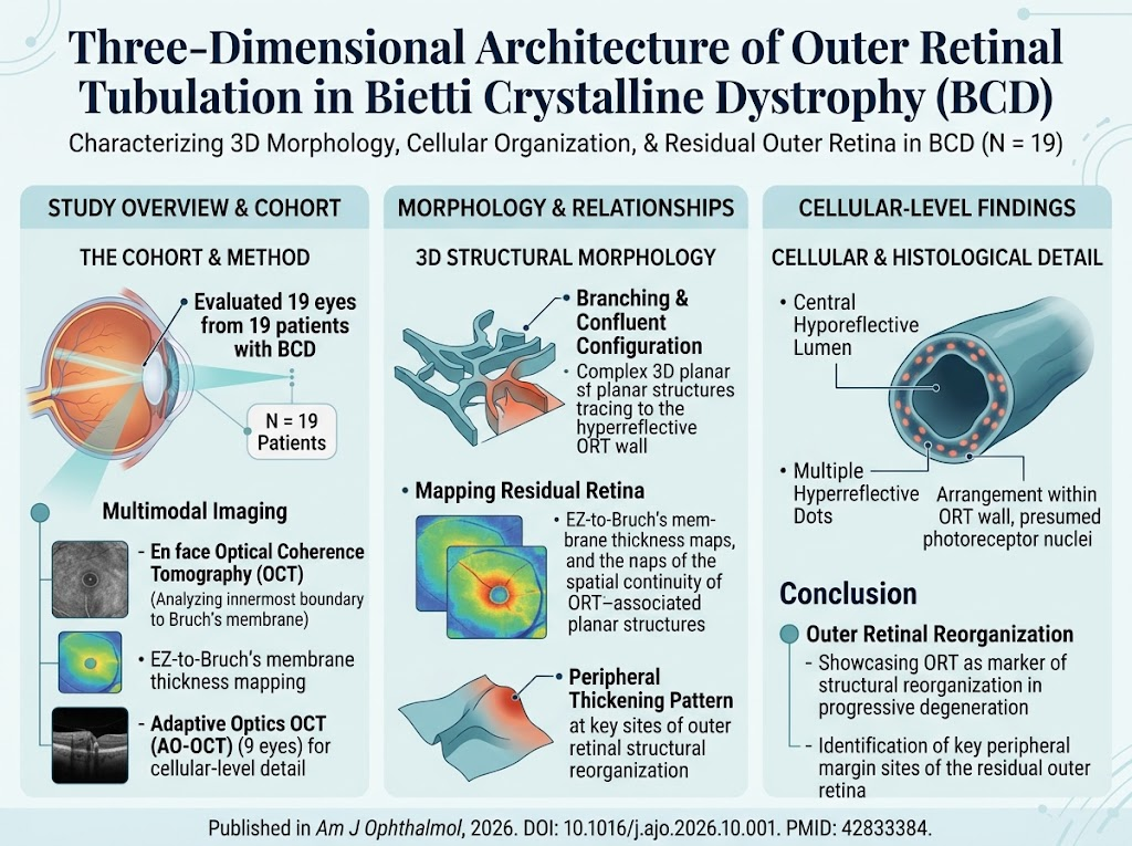
- Outer retinal tubulation (ORT) in Bietti crystalline dystrophy (BCD) exhibits complex, three-dimensional planar configurations involving branching and confluent patterns.
- En face OCT and EZ-to-Bruch’s membrane thickness maps demonstrate spatial association between ORT structures and residual outer retina, highlighting peripheral thickening areas potentially critical for ORT formation.
- Adaptive optics OCT revealed rosette-like photoreceptor nuclei arrangements around hyporeflective lumens within ORT walls, indicating preserved cellular organization amidst degeneration.
- Findings support that ORT represents structural reorganization within the outer retina during progressive BCD degeneration, providing insights for future therapeutic targeting and imaging biomarkers.
Study Background
Bietti crystalline dystrophy (BCD) is a rare inherited retinal disorder characterized by crystalline deposits in the retina and progressive chorioretinal degeneration resulting in vision loss. The natural history involves progressive atrophy chiefly affecting the outer retinal layers, including photoreceptors and retinal pigment epithelium (RPE). Outer retinal tubulation (ORT)—tubular structures seen in degenerative retinal diseases—is increasingly recognized as a morphological hallmark reflecting outer retinal remodeling under stress or injury. Despite its clinical relevance, the precise three-dimensional architecture and cellular organization of ORT in BCD remain poorly characterized. Understanding ORT morphology and its spatial relationship with residual outer retina is critical to unravel the mechanisms underlying retinal remodeling in BCD, potentially informing prognosis and therapeutic strategies.
Study Design
This observational case series enrolled nineteen patients with genetically or clinically confirmed BCD exhibiting outer retinal tubulation. High-resolution imaging modalities were employed to characterize ORT structure:
– En face optical coherence tomography (OCT) images capturing the innermost ORT boundary to Bruch’s membrane to render three-dimensional morphologies.
– Ellipsoid zone (EZ)-like band-to-Bruch’s membrane thickness maps (EZ-to-Bruch’s membrane thickness maps) to quantify outer retinal thickness distribution adjacent to ORT.
– Adaptive optics OCT (AO-OCT) imaging to assess cellular-level organization of ORT walls, focusing on photoreceptor nuclei and luminal structures.
Analysis focused on delineating ORT three-dimensional morphology, residual outer retina relationships, and microstructural cellular patterns within ORT walls.
Key Findings
Out of the nineteen eyes assessed, fifteen provided analyzable en face OCT images and nine yielded interpretable AO-OCT data.
Three-Dimensional Morphology of ORT
En face OCT imaging revealed that ORT manifests as complex planar structures featuring branching and confluent tubulations rather than simple cylindrical forms. The tubular formations were spatially continuous with the EZ-like band, confirming the anatomical linkage between ORT and residual photoreceptor structures. This challenges traditional views of ORT as isolated cystic spaces, instead suggesting an intricate reorganization of outer retinal layers.
EZ-to-Bruch’s Membrane Thickness and Spatial Correlation
EZ-to-Bruch’s membrane thickness maps corresponded closely to ORT-associated planar structures, implying these thickness metrics effectively represent residual outer retinal geometry in the context of BCD. Most eyes demonstrated peripheral thickening of EZ-to-Bruch’s membrane thickness, which, upon quantitative thickness-distance analysis, displayed characteristic peripheral peak patterns particularly in eyes with intermediate-sized ORT areas. These thickened peripheral zones likely represent early or active sites of structural remodeling and ORT genesis.
Cellular-Level Organization via AO-OCT
Adaptive optics OCT revealed multiple hyperreflective dots arranged in rosette-like patterns circumscribing a central hyporeflective lumen within the ORT walls. These dots are consistent with preserved photoreceptor nuclei, suggesting that ORT contains reorganized yet viable photoreceptor elements rather than purely degenerative debris. This cellular architecture supports the hypothesis that ORT formation reflects an adaptive, spatially ordered reconfiguration of photoreceptors during progressive outer retinal degeneration.
Expert Commentary
This comprehensive imaging study significantly advances the understanding of ORT in BCD by elucidating its complex three-dimensional morphology and cellular constituents. The spatial continuity between the EZ-like band and ORT identified via en face OCT supports the concept of ORT as a dynamic structural adaptation to progressive photoreceptor and RPE loss rather than a fixed degenerative endpoint. The peripheral thickening areas mapped by EZ-to-Bruch’s membrane thickness analyses may serve as critical landmarks for monitoring disease progression and therapeutic response.
The AO-OCT findings of photoreceptor nuclei preservation inside ORT tubular walls provide new evidence that ORT involves cellular-level reorganization rather than total cell loss, highlighting potential windows for retina-sparing or restorative interventions. However, given the observational design and limited sample size, further longitudinal studies correlating ORT morphology with functional outcomes are warranted.
Conclusion
Outer retinal tubulation in Bietti crystalline dystrophy represents a complex three-dimensional structural reorganization involving branching planar tubulations contiguous with residual outer retina. Peripheral regions of outer retinal thickening, visualized through thickness mapping, may mark key sites of ORT initiation and progressive remodeling. Cellular-level imaging confirms that photoreceptor nuclei are organized in rosette-like arrays within ORT walls, underscoring the concept of ORT as an adaptive rather than purely degenerative retinal response. These insights into ORT architecture have meaningful implications for understanding disease mechanisms in BCD and potentially guiding future therapeutic strategies to preserve photoreceptor integrity and visual function.
Funding and Clinical Trials
The study was conducted as an observational case series; specific funding sources were not detailed. No clinical trials were referenced.
References
1. Ogata M, Kogo T, Ikeda HO, et al. Three-dimensional architecture of outer retinal tubulation in Bietti crystalline dystrophy. Am J Ophthalmol. 2026 Oct 5; Available from: https://pubmed.ncbi.nlm.nih.gov/42833384/
2. Freund KB, Sadda S, Sarraf D. Outer retinal tubulation: clinical and histologic perspectives. Retina. 2011;31(6):1037–1045.
3. Dolz-Marco R, Freund KB, Curcio CA, Mullins RF. Outer retinal tubulation is part of the photoreceptor degeneration in age-related macular degeneration. Invest Ophthalmol Vis Sci. 2017;58(13):6833–6844.
4. Boon CJ, Klevering BJ, Keunen JE, Hoyng CB, Theelen T, den Hollander AI, et al. Outer retinal tubulation in advanced age-related macular degeneration: implications for retinal remodeling and disease pathophysiology. Eye. 2013;27(8):1332-1340.