Highlights
- Establishment of a fully humanized iPSC-based co-culture model to study interactions between macrophages and sensory neurons.
- Discovery that iPSC-derived macrophages (iMacs) undergo specific morphological and secretory changes in direct response to the injury state of sensory neurons.
- Evidence that macrophages directly amplify spontaneous firing in damaged sensory neurons, providing a mechanistic link between innate immunity and neuropathic pain.
- Identification of neuro-immune signaling pathways as high-priority targets for the development of next-generation non-opioid analgesics.
Background: The Clinical Burden of Neuropathic Pain
Neuropathic pain, arising from lesions or diseases affecting the somatosensory nervous system, remains one of the most challenging conditions to manage in modern clinical practice. Affecting approximately 7% to 10% of the general population, it is characterized by symptoms such as allodynia, hyperalgesia, and—most debilitatingly—spontaneous burning or shooting sensations. Current pharmacological interventions, including gabapentinoids, tricyclic antidepressants, and serotonin-norepinephrine reuptake inhibitors, often yield suboptimal results, with many patients failing to achieve even a 50% reduction in pain intensity while suffering from dose-limiting side effects.The transition from acute injury to chronic pain is driven by maladaptive plasticity within the peripheral and central nervous systems. While historically viewed as a purely neuronal phenomenon, emerging evidence from animal models has increasingly pointed toward the innate immune system, particularly peripheral macrophages, as critical mediators of this process. However, the translation of these findings to human patients has been hindered by the lack of human-specific models that can recapitulate the complex bidirectional signaling between immune cells and neurons. The study by Chrysostomidou et al. addresses this gap by utilizing induced pluripotent stem cell (iPSC) technology to explore these interactions in a fully human system.
Study Design: A Humanized Co-Culture Approach
The research team developed a sophisticated experimental model utilizing human iPSC-derived sensory neurons (iSNs) and iPSC-derived macrophages (iMacs). This approach bypasses the limitations of rodent models, which often fail to replicate human nociceptive signaling and immune responses.
Cellular Differentiation and Characterization
The researchers differentiated iPSCs into sensory neurons expressing classical markers such as BRN3A and peripherin, and into macrophages that exhibited typical phagocytic capabilities and surface markers (CD14, CD68). The iMacs were validated to ensure they reflected the functional characteristics of primary human macrophages.
Injury Modeling and Co-culture
To simulate neuropathic conditions, the iSNs were subjected to specific injury protocols. The iMacs were then introduced into the culture environment. The study utilized a combination of transcriptomic profiling (RNA-seq), secretome analysis, and high-resolution electrophysiology to monitor the phenotypic shifts in both cell types. A key aspect of the design was the comparison between macrophages cultured with healthy neurons versus those cultured with injured neurons, allowing for the isolation of injury-specific signaling.
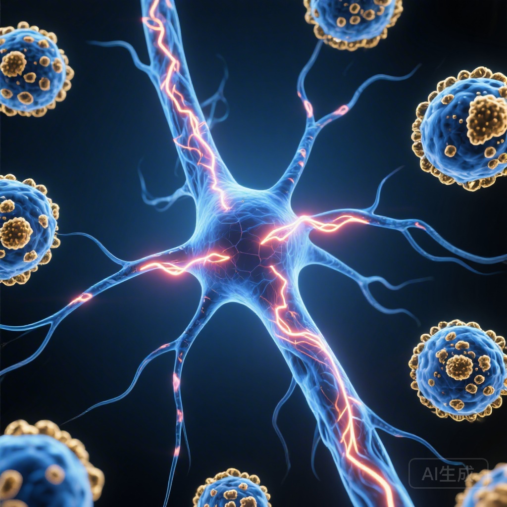
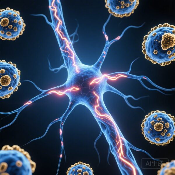
Key Findings: The Bidirectional Dialogue
The results of the study provide compelling evidence that macrophages and sensory neurons exist in a state of dynamic, reciprocal communication.
Macrophage Plasticity is Governed by Neuronal State
One of the most significant findings was that iMacs are highly sensitive to the physiological state of neighboring neurons. When co-cultured with injured iSNs, the macrophages underwent profound changes:
**Morphological Transformation:** iMacs shifted from a surveillance-like state to a more amoeboid, activated morphology.
**Transcriptomic Reprogramming:** Gene expression analysis revealed an upregulation of pro-inflammatory cytokines, chemokines, and growth factors similar to those observed in human nerve biopsy samples from patients with chronic pain.
**Secretory Profile:** The iMacs began secreting a specific milieu of factors that are known to modulate neuronal excitability.
Macrophages as Amplifiers of Spontaneous Firing
The most clinically relevant discovery was the direct effect of these activated iMacs on neuronal function. Spontaneous activity in sensory neurons—firing in the absence of an external stimulus—is a primary driver of the persistent pain felt by patients. The study found that:
Injured neurons already exhibited a baseline increase in spontaneous activity.
The addition of iMacs to the injured neuronal culture significantly amplified this activity, increasing both the frequency and the number of neurons exhibiting spontaneous firing.
Interestingly, iMacs did not induce spontaneous firing in healthy neurons to the same degree, suggesting that a ‘priming’ injury to the neuron is necessary for this pathological neuro-immune feedback loop to take hold.
Expert Commentary: Mechanistic Insights and Clinical Implications
The ability of macrophages to directly modulate the electrophysiological properties of human sensory neurons represents a paradigm shift in our understanding of neuropathic pain pathogenesis. Traditionally, macrophages were viewed primarily as ‘scavengers’ that cleared debris following nerve injury. This study elevates them to the role of active modulators of sensory processing.
Biological Plausibility
Mechanistically, the factors secreted by iMacs (such as TNF-alpha, IL-1beta, and NGF) are known to bind to receptors on the neuronal membrane, leading to the phosphorylation of ion channels like Nav1.7, Nav1.8, and TRPV1. This lowers the threshold for action potential generation and promotes the repetitive firing observed in the study. The humanized nature of this model confirms that these pathways, previously identified in rodents, are indeed operative and potent in human cells.
Study Limitations and Future Directions
While the iPSC model is a significant advancement, it is not without limitations. These cultures lack the structural complexity of an intact nerve, including the presence of Schwann cells and the physical barriers of the epineurium. Additionally, iPSC-derived cells often represent a more fetal-like state rather than mature adult tissue. Future research should aim to incorporate these additional cell types and explore the long-term ‘memory’ of these immune-neuronal interactions to understand why pain persists long after the initial injury has healed.
Conclusion: A New Frontier for Analgesic Development
The study by Chrysostomidou et al. provides a clear mandate for the pharmaceutical industry: to treat neuropathic pain effectively, we must look beyond the neuron. By demonstrating that human macrophages directly amplify the spontaneous neuronal activity that characterizes chronic pain, this research highlights a specific, targetable interface.Disrupting the pathological signaling between macrophages and damaged sensory neurons offers a promising strategy for developing analgesics that are both more effective and have fewer central nervous system side effects than current therapies. As we move toward a more personalized approach to pain management, the human iPSC co-culture model will likely serve as a vital platform for screening compounds that can silence this “immune-driven” neuronal hyperactivity.
References
1. Chrysostomidou P, Hore Z, Somma D, et al. Macrophages amplify spontaneous activity of damaged sensory neurons in a human co-culture model of neuropathic pain. *Brain*. 2026; PMID: 41793058.
2. Scholz J, Woolf CJ. The neuropathic pain triad: neurons, immune cells and glia. *Nat Neurosci*. 2007;10(11):1361-1368.
3. Ghasemlou N, Chiu IM, Julien JP, Woolf CJ. CD11b+ cells mediate the propagation of peripheral inflammation and hyperalgesia following nerve injury. *Pain*. 2015;156(6):1159-1172.
4. Grace PM, Hutchinson MR, Maier SF, Watkins LR. Pathological pain and the neuroimmune interface. *Nat Rev Immunol*. 2014;14(4):217-231.