Highlight
- Baseline EOAD-signature cortical atrophy significantly predicts progression from mild cognitive impairment (MCI) to dementia in early-onset Alzheimer disease (EOAD).
- EOAD-signature atrophy provides prognostic value beyond baseline clinical dementia severity as measured by the Clinical Dementia Rating.
- MRI-based EOAD-signature atrophy has potential utility for individualized prognosis and optimizing clinical trial stratification in EOAD populations.
Study Background
Early-onset Alzheimer disease (EOAD), defined as Alzheimer disease presenting before age 65, represents a clinically heterogeneous syndrome with often more aggressive progression compared to late-onset Alzheimer disease (LOAD). EOAD patients frequently show atypical clinical presentations and differing pathological profiles, demanding biomarkers tailored to this subgroup to improve early diagnosis and prognostic accuracy. Currently, reliable biomarkers to forecast the transition from mild cognitive impairment (MCI), often the prodromal phase of EOAD, to overt dementia are limited. While structural magnetic resonance imaging (MRI) measures have demonstrated prognostic relevance in LOAD, their application for EOAD remains less explored. There is a critical unmet need to identify and validate biomarkers specific to EOAD that can guide clinical decision-making and enrich clinical trials targeting this population.
Study Design
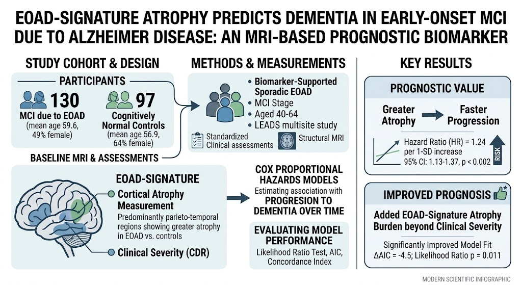
This investigation analyzed patients enrolled in the Longitudinal Early-Onset Alzheimer’s Disease Study (LEADS), a large multisite natural history cohort. Participants aged 40 to 64 years with biomarker-supported sporadic EOAD at the MCI stage underwent standardized clinical evaluations and baseline structural MRI. The EOAD-signature atrophy metric, reflecting cortical thinning in a set of predominantly parieto-temporal regions shown previously to be preferentially affected in EOAD, was quantified from MRI scans. Clinical severity at baseline was assessed using the global Clinical Dementia Rating (CDR) scale. The primary endpoint was progression to dementia during follow-up. Cox proportional hazards models estimated the relationship between baseline EOAD-signature atrophy burden and hazard of progression. Prognostic performance improvement over clinical severity alone was assessed using likelihood ratio tests, Akaike Information Criterion (AIC), and Harrell’s concordance index.
Key Findings
The study included 130 individuals with MCI due to EOAD (mean age 59.6 years, 49% female) and 97 cognitively normal controls (mean age 56.9 years, 64% female). Baseline EOAD-signature atrophy was significantly elevated in MCI patients compared to controls. Importantly, greater baseline cortical atrophy predicted a faster progression to dementia, with a hazard ratio (HR) of 1.24 per 1-standard deviation (SD) increase in atrophy (95% CI: 1.13–1.37, p < 0.002). When combined with baseline clinical dementia severity, the MRI measure significantly improved prognostic model fit (ΔAIC = -4.5; likelihood ratio test p = 0.011), indicating that EOAD-signature atrophy added prognostic value beyond clinical assessment alone. Harrell’s concordance index also improved, suggesting enhanced prediction accuracy.
No safety concerns were reported as this was an observational imaging study. The effect size indicates a meaningful increase in risk stratification capability when incorporating structural MRI-based atrophy measures.
Expert Commentary
This study addresses a critical knowledge gap in EOAD biomarker research by demonstrating that a disease-specific cortical atrophy signature measured via MRI can robustly predict dementia progression from MCI. The EOAD-signature focuses on parieto-temporal regions that are differentially vulnerable in EOAD, contrasting with the predominantly temporoparietal atrophy seen in LOAD. This biologically plausible pattern strengthens confidence in the marker’s validity and specificity.
Although the sample size is modest, the multisite design and biomarker confirmation increase generalizability. Potential limitations include the sample’s selection bias as participants were enrolled in a natural history study and may not fully represent community populations. Furthermore, longitudinal MRI data were not detailed in this study and could further refine prognostication models.
Future research should explore combining EOAD-signature atrophy with other biomarkers such as tau PET or cerebrospinal fluid markers to enhance predictive power. Integration of these tools into clinical workflows could allow earlier and more personalized intervention planning.
Conclusion
Baseline cortical atrophy within an EOAD-specific parieto-temporal signature measured by structural MRI predicts progression from MCI to dementia in early-onset Alzheimer disease. This MRI biomarker provides prognostic information beyond clinical severity assessments and holds promise for clinical application in individualized prognosis and clinical trial stratification. These findings underscore the value of disease-specific imaging signatures in refining diagnosis and prognosis in neurodegenerative disorders.
Funding and Clinical Trials Registry
The LEADS Consortium study was supported by grants from the National Institutes of Health (NIH) and affiliated academic institutions. Detailed funding sources and trial registration information can be accessed via the original publication and the LEADS study protocol.
References
1. Paranhos T, Katsumi Y, Brickhouse MJ, et al. EOAD-Signature Atrophy Predicts Dementia in Early-Onset MCI due to Alzheimer Disease: An MRI-Based Prognostic Biomarker. Neurology. 2026 Aug 26;107(7):e2185-219. doi: 10.1212/WNL.0000000000018520. PMID: 42647766.
2. La Joie R, Visani AV, Baker SL, et al. Multicohort study of longitudinal amyloid and tau PET in preclinical and early Alzheimer’s disease. Neurology. 2020;95(11):e1334-e1346.
3. Jack CR Jr, Knopman DS, Jagust WJ, et al. Tracking pathophysiological processes in Alzheimer’s disease: an updated hypothetical model of dynamic biomarkers. Lancet Neurol. 2013 Feb;12(2):207-16.
4. Ossenkoppele R, Schonhaut DR, Schöll M, et al. Tau PET patterns mirror clinical and neuroanatomical variability in Alzheimer’s disease. Brain. 2016 Nov;139(Pt 5):1551-1567.
5. Dubois B, Feldman HH, Jacova C, et al. Advancing research diagnostic criteria for Alzheimer’s disease: the IWG-2 criteria. Lancet Neurol. 2014 Jun;13(6):614-29.