Highlight
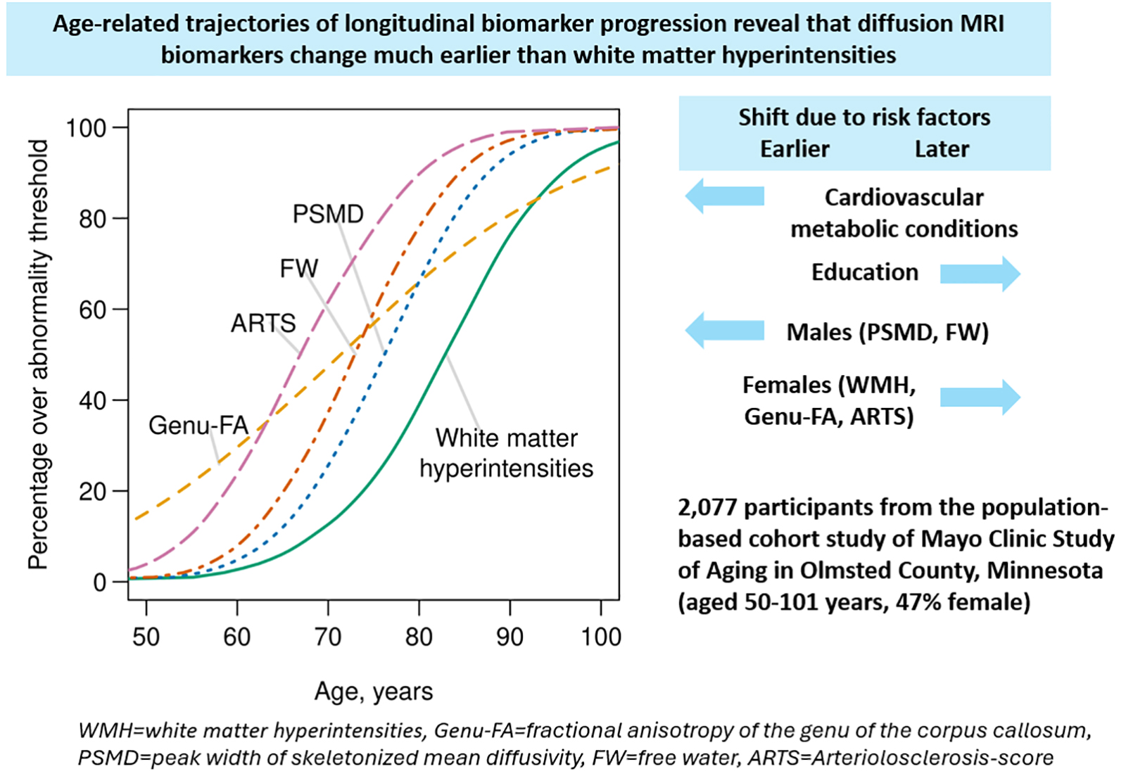
This study demonstrates that diffusion MRI biomarkers detect cerebral small vessel disease (CSVD) microstructural abnormalities at least a decade before traditional white matter hyperintensities (WMH) become apparent on fluid-attenuated inversion recovery (FLAIR) MRI. The findings reveal a prolonged pre-WMH phase characterized by abnormal fractional anisotropy, mean diffusivity, free water, and a novel arteriolosclerosis score. Cardiometabolic conditions and sex influence the timing and progression of these biomarkers, underscoring opportunities for earlier diagnosis and targeted prevention.
Study Background
Cerebral small vessel disease is a leading cause of vascular cognitive impairment and stroke, primarily detected by white matter hyperintensities (WMH) on conventional MRI. However, WMH represent relatively late-stage tissue injury. Earlier microstructural alterations likely precede visible WMH and may offer a window for preventive interventions. Diffusion MRI provides sensitive biomarkers of white matter integrity, including fractional anisotropy (FA), mean diffusivity (MD), free water content, and novel automated imaging markers correlated to small vessel pathology such as the arteriolosclerosis score. Despite growing interest, the temporal evolution of these diffusion metrics in relation to WMH onset and their modulation by vascular risk factors remains poorly characterized in large aging cohorts.
Study Design
The Mayo Clinic Study of Aging prospectively enrolled 2,077 participants aged 50 to 101 years from Olmsted County, Minnesota, with serial neuroimaging data collected from May 2005 through September 2024. Inclusion criteria required at least two longitudinal FLAIR and diffusion MRI scans per participant. Researchers applied multioutput nonlinear mixed-effects models to delineate temporal trajectories of WMH volume and four diffusion MRI biomarkers: fractional anisotropy of the genu of the corpus callosum (FA-Genu), peak width of skeletonized mean diffusivity (PSMD), free water, and an automated arteriolosclerosis score. Models accounted for participant-specific timing shifts, biomarker correlations, and the influence of sex, education, APOE ε4 genotype, and cardiometabolic risk factors (such as hypertension, diabetes, and hyperlipidemia).
Key Findings
The average baseline age was 78 years; 47% were female, 28% carried the APOE ε4 allele, and 80% were cognitively unimpaired. Follow-up averaged 5.2 years for FLAIR MRI and 4.3 years for diffusion MRI. Key results included:
- Early Biomarker Abnormalities: The arteriolosclerosis score became abnormal in 50% of participants 16 years before WMH onset, FA-Genu abnormality preceded WMH by 12 years, free water by 10 years, and PSMD by 7 years (with narrow confidence intervals under one year), indicating a prolonged preclinical phase of CSVD.
- Correlated Pathology: Global markers of white matter injury (arteriolosclerosis score, free water, PSMD, WMH) were intercorrelated, suggesting these diffusion abnormalities and WMHs reflect shared substrates of vascular injury.
- Distinctiveness of FA-Genu: FA-Genu was only weakly coupled with WMH, indicating FA-Genu abnormalities may capture vascular microstructural injury progressing more linearly and continuously throughout adulthood, rather than abrupt late-stage damage.
- Influence of Cardiometabolic Risk: Participants with cardiometabolic conditions exhibited earlier and accelerated progression across all imaging biomarkers, reinforcing the systemic vascular contributions to CSVD.
- Sex Differences: Women showed earlier abnormalities in WMH, FA-Genu, and arteriolosclerosis score, while men exhibited earlier PSMD and free water changes, highlighting sex-specific vulnerability or pathophysiology.
Expert Commentary
This landmark longitudinal study provides compelling evidence that diffusion MRI biomarkers can identify small vessel disease-associated brain changes long before conventional WMHs emerge. The identification of a prolonged pre-WMH phase lasting up to 16 years offers a crucial diagnostic window for cerebrovascular risk modification and secondary prevention. The integration of multiple diffusion metrics and an automated arteriolosclerosis score strengthens the analysis by capturing different facets of microvascular brain injury.
Of particular note, FA-Genu’s weak coupling with WMH and its more linear decline suggest it may serve as an independent marker of ongoing vascular injury, potentially useful in monitoring disease trajectory or response to interventions. The observed sex differences and impact of cardiometabolic risk factors underscore the importance of personalized risk profiling in CSVD management.
Limitations include the observational nature restricting causal inference and potential cohort-specific factors related to Olmsted County demographics that may limit generalizability. Furthermore, while diffusion MRI biomarkers offer enhanced sensitivity, their specificity to small vessel pathology versus other neurodegenerative processes warrants further validation.
Conclusion
This study redefines our understanding of the temporal progression of cerebral small vessel disease by establishing that microstructural white matter injury detectable via diffusion MRI precedes WMH by at least a decade. Early diffusion MRI biomarkers and the arteriolosclerosis score hold promise to serve as sensitive early indicators to stratify risk, guide therapeutic interventions, and potentially prevent progression to symptomatic CSVD and vascular cognitive impairment.
Future research should focus on validating these diffusion MRI biomarkers in diverse populations, exploring underlying pathophysiologic mechanisms, and integrating them into clinical trials aimed at early CSVD intervention.
Funding and ClinicalTrials.gov
The study was supported by the Mayo Clinic, National Institutes of Health grants, and other institutional funding. Specific trial registration numbers are not reported.
References
- Vemuri P, Hu M, Lundt ES, et al. Diffusion MRI Identifies a Prolonged Pre-WMH Phase in Cerebral Small Vessel Disease. Stroke. 2026;57(9):2789-2800. PMID: 42483814.
- Wardlaw JM, Smith C, Dichgans M. Small vessel disease: mechanisms and clinical implications. The Lancet Neurology. 2019;18(7):684-696.
- Maillard P, Seshadri S, Beiser A, et al. White matter lesion progression: a clinical marker of small vessel disease progression. Neurology. 2012;79(8):792-798.
- O’Sullivan M, Craggs LJ, Barker GJ, et al. Patterns of cerebral white matter damage detected using diffusion tensor imaging in CADASIL: a genetic form of small vessel disease. Neurology. 2001;56(10):1277-1284.