 Highlight
- Contemporary UGH syndrome predominantly involves posterior chamber intraocular lenses (IOLs), especially those positioned in the sulcus.
- The median interval from causative surgery to UGH diagnosis is nearly 5 years, with late-onset cases (>10 years) showing distinct clinical features including pseudophacodonesis and Soemmering ring formation.
- Nearly half of eyes experience recurrent disease, and persistent ocular hypertension remains common despite treatment interventions.
- Surgical intervention is frequently necessary, resulting in improved intraocular pressure (IOP) and best-corrected visual acuity (BCVA) outcomes at 12 months.
Study Background
Uveitis-glaucoma-hyphema (UGH) syndrome is a well-recognized but relatively infrequent complication associated with intraocular lens (IOL) implantation. Traditionally linked to malpositioned or incompatible lens implants, UGH syndrome involves chronic inflammation (uveitis), elevated intraocular pressure (glaucoma), and intraocular bleeding (hyphema), which can jeopardize vision. Despite advances in cataract surgery and IOL design, UGH syndrome persists as a diagnostic and therapeutic challenge, particularly with late-onset presentations. Understanding modern clinical presentations, risk factors, and outcomes is critical for optimizing patient care and preserving vision in affected individuals.
Study Design
This retrospective clinical cohort study reviewed 232 eyes from 226 patients diagnosed with UGH syndrome at a tertiary academic referral center between October 2015 and December 2025. The study extracted demographic information, details on IOL type and positioning, ocular clinical and imaging findings, management strategies employed, and outcomes related to visual acuity and IOP control. The cohort was stratified by timing of UGH onset from causative surgery into early-onset (≤1 year), intermediate (1-10 years), and late-onset (>10 years) groups to elucidate temporal patterns.
Key Findings
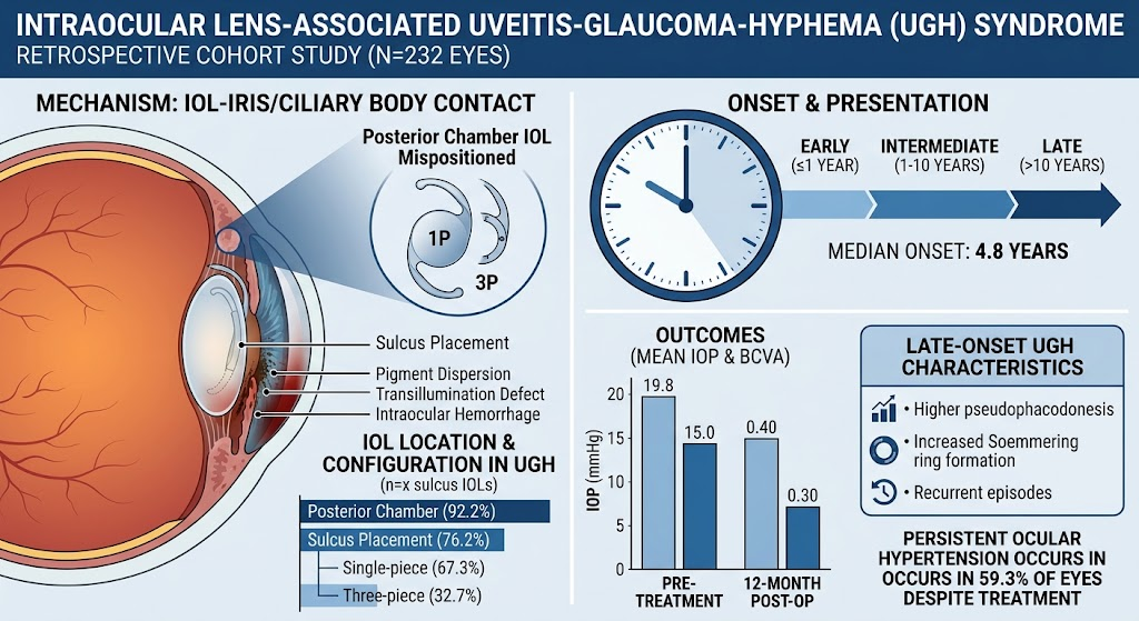
The cohort had a mean age of 68.4 years at diagnosis. Posterior chamber IOLs accounted for the vast majority (92.2%) of cases, with most lenses located in the sulcus (76.2%). Among sulcus-positioned lenses, two-thirds were single-piece and one-third three-piece IOL configurations.
The median time from surgery to UGH diagnosis was 4.8 years, indicating a propensity for delayed onset. Clinical signs most commonly included IOL contact with the iris or ciliary body (77.3%), pigment dispersion (59.3%), iris transillumination defects (50.0%), and intraocular hemorrhage (48.7%). Recurrent UGH episodes were seen in nearly half (46.2%) of eyes, highlighting the chronic and relapsing nature of the syndrome.
Surgical treatment, such as IOL repositioning or exchange, was undertaken in 77.2% of eyes. Following intervention, the median IOP decreased significantly from 19.8 mmHg pre-treatment to 15.0 mmHg at 12 months, and median BCVA improved modestly from 0.40 to 0.30 logMAR, reflecting functional visual gains.
Persistent ocular hypertension despite treatment was prevalent, occurring in 59.3% of eyes. Late-onset disease uniquely exhibited higher rates of pseudophacodonesis—indicative of lens instability—and Soemmering ring formation, commonly associated with recurrent symptomatic episodes.
Expert Commentary
These findings reflect the evolving clinical spectrum of UGH syndrome in the era of modern IOLs, with the majority of cases arising from posterior chamber lenses placed in the sulcus, a position known to predispose to iris chafing and pigment dispersion if lens design or sizing is suboptimal. The significant prevalence of late-onset UGH underscores the necessity for long-term vigilance in post-cataract patients presenting with inflammation or elevated IOP.
Persistent ocular hypertension despite surgical and medical treatment remains a management challenge, emphasizing the importance of early detection and tailored interventions. The association of pseudophacodonesis and Soemmering rings with late-onset disease suggests a pathophysiologic link involving lens instability and residual capsular material that may perpetuate iris trauma and inflammation.
Limitations of the study include its retrospective nature and single-center setting, which might limit generalizability. Nonetheless, the large cohort and detailed stratification by onset timing contribute valuable clinical insights.
Conclusion
Intraocular lens-associated UGH syndrome is predominantly a late complication of posterior chamber IOL implantation, particularly in lenses placed in the sulcus. Clinical manifestations commonly include iris or ciliary body contact and pigment dispersion, which lead to recurrent inflammation, hyphema, and secondary glaucoma. Surgical management improves visual and IOP outcomes but does not preclude persistent ocular hypertension, especially in late-onset cases characterized by lens instability and Soemmering ring formation.
Ophthalmologists should maintain a high index of suspicion for UGH syndrome in patients with unexplained uveitis and glaucoma following cataract surgery, regardless of the elapsed time since surgery. Timely diagnosis and appropriate surgical intervention are vital to prevent irreversible ocular damage and optimize long-term visual outcomes. Further prospective studies are needed to establish standardized management guidelines and explore preventive strategies targeting sulcus-placed IOLs.
Reference
Kang S, Theotoka D, Ballouz D, Chen JA, Zhu AY, Smiddy WE, Gedde SJ, Bitrian E, Townsend JH, Flynn HW Jr, Yoo SH. Intraocular Lens-Associated Uveitis-Glaucoma-Hyphema Syndrome. Am J Ophthalmol. 2026 Aug 13:S0002-9394(26)00466-6. doi: 10.1016/j.ajo.2026.08.016. Epub ahead of print. PMID: 42595287.