Highlight
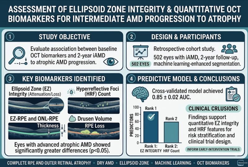
- Ellipsoid zone (EZ) integrity metrics and hyperreflective foci (HRF) counts are strong baseline predictors of intermediate AMD progression to advanced atrophic AMD over 2 years.
- Machine learning-enhanced quantitative OCT analysis effectively identifies subtle retinal structural changes not apparent on conventional imaging.
- A predictive random forest model using multiple OCT biomarkers achieved high accuracy (AUC = 0.85) in forecasting disease progression.
- These findings underscore the potential for early risk stratification, informing clinical trial design and targeted therapeutic interventions to delay atrophic AMD onset.

Study Background
Age-related macular degeneration (AMD) is a leading cause of irreversible vision loss in older adults globally. Intermediate AMD (iAMD) represents a critical stage wherein structural retinal changes are evident but before irreversible atrophy ensues. Current clinical challenges include identifying which patients with iAMD are at higher risk of rapid progression to advanced atrophic AMD, characterized by retinal pigment epithelium (RPE) loss and outer retinal atrophy, as these patients could benefit most from early intervention. Optical coherence tomography (OCT) offers high-resolution imaging of the retinal layers, allowing for detailed structural assessment. Among various OCT features, the integrity of the ellipsoid zone (EZ)—representing the photoreceptor inner segment/outer segment junction—is postulated to reflect photoreceptor health, yet its quantitative role in predicting atrophic progression remains incompletely characterized. Furthermore, other OCT biomarkers such as drusen volume, hyperreflective foci (HRF), outer nuclear layer thickness, and hypertransmission signals may contribute to risk stratification. This study addresses the unmet need for validated prognostic biomarkers through quantitative and machine learning-augmented OCT analysis to predict iAMD progression over a 2-year period.
Study Design
This retrospective cohort study evaluated 502 eyes diagnosed with intermediate AMD without evidence of atrophy or exudation at baseline. Participants had spectral-domain OCT scans at baseline and 2-year follow-up. A validated, multi-layer segmentation platform enhanced by machine learning automatically quantified multiple retinal structural parameters, including EZ-RPE thickness, partial and total EZ attenuation, drusen volume, HRF counts, the thickness of the outer nuclear layer to RPE (ONL-RPE), and total RPE loss. Certified readers reviewed and corrected segmentations to ensure accuracy.
In addition, fully automated deep-learning models generated specific metrics: hypertransmission areas (indicative of RPE disruption) and ‘EZ at-risk’ zones (regions of abnormal EZ-RPE thinning without concurrent RPE loss). A random forest classifier was developed using baseline OCT features to predict 2-year progression to advanced atrophic AMD—defined by OCT criteria of complete RPE and outer retinal atrophy (cRORA) with lesion area ≥0.05 mm2. Model performance underwent robust assessment via 5-fold stratified cross-validation.
Key Findings
Among the 502 eyes analyzed, those progressing to advanced atrophic AMD at two years exhibited significant baseline differences compared to non-converters. Specifically, converters showed:
– Greater degrees of partial and total EZ attenuation, indicating compromised photoreceptor integrity.
– Reduced EZ-RPE and ONL-RPE thickness measurements, reflecting thinning of vital retinal layers.
– Elevated drusen volume, consistent with higher pathological burden.
– Increased HRF counts, suggestive of migrating RPE cells or inflammatory processes.
– Higher deep-learning-derived hypertransmission areas and expanded EZ at-risk regions.
Statistical significance was consistently strong across these parameters (all p-values <0.05).
The predictive random forest model integrating all baseline OCT features achieved a mean area-under-the-curve (AUC) of 0.85 ± 0.02, representing high discriminatory ability. Feature importance analysis ranked EZ integrity measures (attenuation metrics and thicknesses) and HRF count as the most influential determinants of future atrophic progression. Notably, this model outperformed simpler univariate predictors and demonstrated the added value of combining multiple quantitative biomarkers.
Expert Commentary
The findings by Matar et al. elucidate the critical role of ellipsoid zone integrity and hyperreflective foci in the pathophysiology and prognostication of AMD progression. The ellipsoid zone, representing mitochondrial-rich photoreceptor segments, acts as a surrogate marker for photoreceptor viability. Its attenuation or loss is a direct reflection of photoreceptor dysfunction and impending atrophy. HRF, which may represent activated microglia or displaced RPE cells, further highlights ongoing retinal stress and remodeling.
The leverage of machine learning for multilayer segmentation and automated biomarker quantification addresses inherent variability and enhances reproducibility, critical for both clinical practice and research. Furthermore, the creation of composite predictive models heralds a move towards personalized risk assessment, potentially guiding clinical decision-making regarding surveillance intervals and therapeutic trial enrollment.
However, limitations inherent to retrospective analyses, including selection bias and the lack of standardized systemic or demographic adjustments, must be acknowledged. Additionally, external validation in diverse populations and incorporation of functional measures such as visual acuity or patient-reported outcomes would enrich the clinical applicability.
Conclusion
This study demonstrates that quantitative OCT biomarkers, especially ellipsoid zone integrity and hyperreflective foci, are robustly associated with 2-year progression from intermediate to advanced atrophic AMD. The use of advanced machine learning techniques enables precise risk stratification and identification of high-risk eyes before clinically overt atrophy. These insights have direct implications for clinical care by enabling early identification of patients suitable for closer monitoring or preventive interventions. Moreover, these biomarkers provide valuable endpoints for designing and evaluating future clinical trials targeting AMD progression. Future research should focus on prospective validation, integration with systemic risk factors, and exploring therapeutic avenues targeting early photoreceptor and RPE preservation.
Funding and Clinical Trials
The study does not specify external funding sources or clinical trial registration. Further details on funding and trial status would be beneficial for transparency.
References
1. Matar K, Delaney A, Indurkar A, et al. Assessment of Ellipsoid Zone Integrity and Other Quantitative OCT Biomarkers for Intermediate AMD Progression to Atrophy. Ophthalmology. 2026 Aug 10. PMID: 42575302.
2. Ferris FL 3rd, Wilkinson CP, Bird A, et al. Clinical classification of age-related macular degeneration. Ophthalmology. 2013 Apr;120(4):844-51.
3. Sadda SR, Keane PA, Ouyang Y, et al. Machine Learning Approaches for the Quantitative Assessment of Retinal Structure Using Optical Coherence Tomography. Ophthalmology. 2020 Jan;127(1):134-150.
4. Fleckenstein M, Fritsche LG, Keilhauer CN, et al. The progression of age-related macular degeneration: Clinical and imaging characteristics. Prog Retin Eye Res. 2021 Mar;82:100896.
This article emphasizes the integration of novel imaging biomarkers and machine learning to enhance early disease risk prediction in AMD, a major step forward in personalized ophthalmic care.