Highlight
This study evaluates the use of hybrid indocyanine green (ICG) and technetium-99m-labeled radiocolloid (99ᵐTc-Nanocoll) for sentinel lymph node biopsy (SLNB) in early-stage vulvar cancer. Key findings include high sentinel node detection rates, especially in midline tumors, and low groin recurrence rates with excellent survival outcomes, supporting the feasibility and oncologic safety of incorporating near-infrared fluorescence imaging.
Study Background
Vulvar cancer, although a relatively rare gynecologic malignancy, poses significant clinical challenges due to its morbidity and impact on quality of life. Lymphatic spread, primarily to the inguinal lymph nodes, is a major prognostic factor influencing treatment and outcomes. Sentinel lymph node biopsy (SLNB) has become the standard of care for early-stage vulvar cancer, reducing the morbidity associated with complete lymphadenectomy by targeting the first draining lymph nodes for pathological evaluation.
The conventional SLNB technique employs technetium-99m-labeled radiocolloid (99ᵐTc-Nanocoll) and blue dye for lymphatic mapping. However, limitations include incomplete identification of sentinel lymph nodes (SLNs), especially in midline tumors where lymphatic drainage may be bilateral or atypical. Indocyanine green (ICG), a near-infrared fluorescent tracer, has emerged as a complementary tool offering real-time intraoperative visualization of lymphatic pathways but lacks extensive data on efficacy and long-term oncologic safety in vulvar cancer.
Study Design
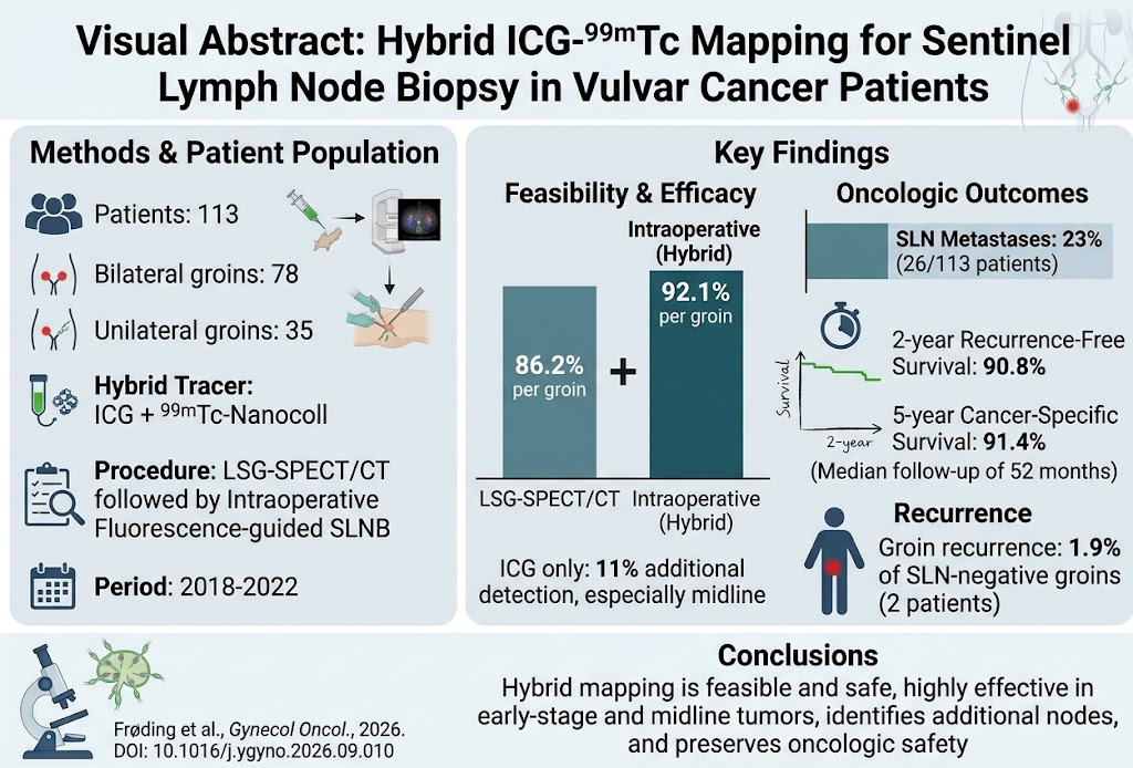
This prospective, consecutive cohort study enrolled 113 patients with early-stage vulvar cancer from February 2018 to August 2022 who underwent SLNB. Both unilateral and bilateral groin assessments were included (35 unilateral and 78 bilateral, totaling 191 groins). All patients underwent preoperative lymphoscintigraphy-single photon emission computed tomography/computed tomography (LSG-SPECT/CT) using 99ᵐTc-Nanocoll to identify sentinel lymph nodes, followed by intraoperative near-infrared fluorescence-guided SLNB using a hybrid tracer combining ICG and 99ᵐTc-Nanocoll.
Endpoints included sentinel lymph node detection rates (preoperative and intraoperative), pathological findings of lymph node metastases, rates of groin recurrence, recurrence-free survival (RFS), and cancer-specific survival (CSS). Statistical analyses employed Kaplan-Meier survival curves and multivariate Cox regression adjusted for cancer stage.
Key Findings
Sentinel Lymph Node Detection: Preoperative LSG-SPECT/CT detected sentinel lymph nodes in 86.2% of groins (94.6% per patient). Intraoperative detection was superior, identifying SLNs in 92.1% of groins and 98.2% of patients overall. Notably, ICG alone identified SLNs in 11% of groins that were not detected by LSG-SPECT/CT, predominantly in cases with midline tumors—a critical area where lymphatic drainage is more complex and bilateral.
Pathological Findings and Recurrence: SLN metastases were found in 26 patients (23%). Over a median follow-up of 52 months (interquartile range 39–67 months), isolated groin recurrence occurred in only 1.9% of SLN-negative groins (2 patients). This low recurrence rate emphasizes the reliability of the hybrid ICG-99ᵐTc approach in accurately staging nodal status.
Survival Outcomes: The two-year recurrence-free survival was 90.8%, with no significant difference between patients whose SLNs were successfully mapped and those with less clear mapping. Cancer-specific survival rates at two and five years were 97.3% and 91.4%, respectively, reflecting excellent long-term outcomes consistent with effective nodal staging and management.
Clinical Implications: The hybrid tracer approach enhanced sentinel node detection beyond standard radiocolloid alone, particularly in midline tumor cases where conventional methods may miss sentinel nodes. The combination of ICG’s real-time fluorescence imaging and the established radiocolloid method offers complementary strengths—real-time surgical guidance and preoperative anatomical localization.
Expert Commentary
Sentinel lymph node biopsy in vulvar cancer is pivotal for personalized surgical management that balances effective oncologic control with minimization of surgical morbidity. The integration of near-infrared fluorescence imaging using ICG into the SLNB protocol represents an important advancement. It offers surgeons enhanced visual guidance, potentially reducing false negatives and improving detection rates especially in challenging anatomical contexts such as midline tumors.
These findings align with emerging clinical data supporting ICG’s utility in other malignancies, such as breast and cervical cancers. However, long-term follow-up and multicenter validation remain essential to confirm oncologic safety across broader clinical cohorts. Additionally, the logistical and resource implications of hybrid tracer use, including availability of fluorescence imaging equipment and tracer preparation, warrant consideration.
Study limitations include its single-cohort non-randomized design, which may introduce selection bias. Nonetheless, the consecutive enrollment and robust follow-up strengthen the validity of conclusions. Future randomized controlled trials comparing hybrid tracer techniques with standard mapping could definitively establish superiority or equivalence.
Conclusion
This study establishes the feasibility, safety, and efficacy of a hybrid mapping technique combining near-infrared fluorescent ICG and 99ᵐTc-Nanocoll for sentinel lymph node biopsy in early-stage vulvar cancer. It enhances sentinel node detection rates, including in anatomically complex midline tumors, without compromising oncologic safety, as reflected in low groin recurrence and high survival rates. Incorporation of real-time fluorescence imaging into clinical practice may optimize surgical staging and personalized management, ultimately improving patient outcomes in this rare and challenging cancer.
Funding and ClinicalTrials.gov
The original publication does not specify funding sources or trial registration numbers. Future studies should ensure transparent reporting of these elements to enhance research rigor and reproducibility.
References
Frøding LP, Christensen A, Kristensen E, Mortensen J, Juhl K, Schnack TH. Feasibility of real-time near-infrared fluorescence tracer imaging in sentinel lymph node biopsy for vulvar cancer patients. Gynecol Oncol. 2026 Sep 21;213:89-94. PMID: 42767171.
Additional relevant references for clinicians may include:
- Levenback CF, Ali S, Coleman RL, et al. The safety of sentinel lymph node biopsy in vulvar cancer: Long-term follow-up results of a prospective multicenter study. J Clin Oncol. 2012;30(6):0549-555.
- van der Zee AGJ, Oonk MH, de Hullu JA, et al. Sentinel node dissection is safe in the treatment of early-stage vulvar cancer. J Clin Oncol. 2008;26(6):884-889.
- Kurumboor Prakash A, Singh SS, Bhosale U, et al. Near-infrared fluorescence imaging using indocyanine green for sentinel lymph node mapping in gynecologic cancers: A systematic review. Gynecol Oncol. 2021;163(3):622-630.