Highlight
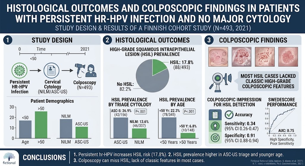
Persistent high-risk HPV infection, even with normal or minor cytological changes, can harbor a considerable prevalence of histologically confirmed high-grade squamous intraepithelial lesions (HSIL). Younger patients and those with atypical squamous cells of undetermined significance (ASC-US) cytology present an elevated risk. Traditional colposcopic assessment features of HSIL are frequently absent in this group, resulting in low sensitivity of colposcopic impressions despite high specificity. The Swedescore system shows moderate performance, indicating the need for adjusted biopsy thresholds in managing this evolving screening population.
Study Background
Cervical cancer screening has evolved from cytology-based methods to primary high-risk human papillomavirus (hrHPV) testing due to the latter’s higher sensitivity for detecting precancerous lesions. However, hrHPV testing possesses lower specificity compared to cytology, leading to an increased number of colposcopy referrals and identification of a new patient subset—those with persistent hrHPV infections but without major cytological abnormalities. This group poses a clinical challenge, as they have the potential risk for high-grade lesions that might be missed under conventional screening algorithms. Understanding the histological outcomes and colposcopic appearances in these patients is critical for optimizing clinical management and preventing progression to cervical cancer.
Study Design
This retrospective cohort study analyzed 493 Finnish women referred for colposcopy in 2021 through the national cervical cancer screening program due to persistence of hrHPV infection accompanied by triage cytology showing either negative for intraepithelial lesion or malignancy (NILM) or atypical squamous cells of undetermined significance (ASC-US). The study described and compared colposcopic findings and histological results across subgroups defined by referral cytology and age. Diagnostic performance of colposcopic impression and systematic scoring by the Swedescore system for detecting HSIL was evaluated using sensitivity, specificity, and receiver operating characteristic (ROC) curve analysis.
Key Findings
The key outcome was the surprisingly high prevalence of histologically confirmed HSIL in this cohort—17.8% overall. Stratification revealed significantly higher HSIL prevalence among women with ASC-US cytology (26.9%) than those with NILM cytology (13.6%). Age was also a significant factor, with women under 50 years old displaying a HSIL prevalence of 22.3%, contrasting with only 6.8% in women older than 50 years.
Interestingly, most HSIL cases did not exhibit classical colposcopic features traditionally associated with high-grade lesions, such as dense acetowhite epithelium, coarse mosaic patterns, or punctation. As a result, the sensitivity of colposcopic impression for HSIL was notably low at 34%, although specificity was high at 91%, indicating that while colposcopy was good at ruling out HSIL when impressions were negative, many lesions remained undetected on visual assessment alone.
The Swedescore system, a structured colposcopic scoring tool, demonstrated moderate ability to discriminate HSIL with an area under the curve (AUC) of 0.75. The score maintained high specificity but suffered from poor sensitivity, reflecting its limited efficacy as a standalone diagnostic tool in this population.
Expert Commentary
The findings presented here underscore the evolving challenges in cervical cancer screening as primary hrHPV testing expands worldwide. Historically, women with normal cytology and low-grade abnormalities might have been considered low-risk and less rigorously evaluated. However, persistent hrHPV infection clearly confers a substantive risk for HSIL development, especially in younger women and those with ASC-US cytology.
The low sensitivity of colposcopy in this context challenges conventional reliance on visual cues alone to guide biopsy decisions. Given the absence of overt high-grade features in many lesions, colposcopists should maintain a lower threshold for performing biopsies, even when colposcopic impressions appear benign. This approach might mitigate underdiagnosis and ensure timely intervention.
Current scoring systems like Swedescore add objectivity but have intrinsic limitations, highlighting the need for improved adjunctive diagnostic tools—such as molecular markers or enhanced imaging techniques—to refine risk stratification and clinical decision-making.
This study’s retrospective design and focus on a single national screening program may limit generalizability, but its large sample size and standardized national protocols confer strength. Future research should explore longitudinal outcomes to clarify the natural history and clinical significance of HSIL detected in this setting and assess the impact of different management strategies on patient outcomes.
Conclusion
The transition to hrHPV-based cervical cancer screening unveils a considerable subset of women with persistent infections who, despite benign or minor cytology, harbor significant high-grade cervical lesions. Age and cytological atypia modulate HSIL risk within this group. Colposcopic evaluation alone, especially reliant on classic visual features, lacks sufficient sensitivity, emphasizing the necessity of cautious biopsy strategies and potential integration of more sensitive diagnostic modalities.
Clinicians should be aware of this nuanced risk landscape, adapt colposcopic interpretations accordingly, and maintain vigilance to prevent overlooked precancerous lesions evolving into invasive disease. Continued investigation is warranted to optimize management algorithms and ensure screening benefits are maximized while minimizing unnecessary procedures.
Funding and Clinical Trials
The study was conducted within the Finnish national cervical cancer screening program framework. No external funding or clinical trial registration information was stated.
References
- Hulmi J, Virtanen A, Sadeluoto H, Tarkkanen J, Kalliala I, Heinonen A. Histological outcomes and colposcopic findings in patients with persistent high-risk human papillomavirus infection without major cytological findings. Am J Obstet Gynecol. 2026 Mar 25;235(2):356-364. PMID: 41895365.
- Cuzick J, Clavel C, Petry KU, et al. Overview of the European and North American studies for HPV testing in primary cervical cancer screening. Int J Cancer. 2006;119(5):1095-1101.
- Katki HA, Kinney WK, Fetterman B, et al. Risk stratification using HPV genotyping and cytology to guide management of HPV-positive women: A prospective cohort study. J Natl Cancer Inst. 2011;103(17):1358-1368.
- Massad LS, Einstein MH, Huh WK, et al. 2012 Updated consensus guidelines for the management of abnormal cervical cancer screening tests and cancer precursors. J Low Genit Tract Dis. 2013;17(5 Suppl 1):S1-S27.