Highlight
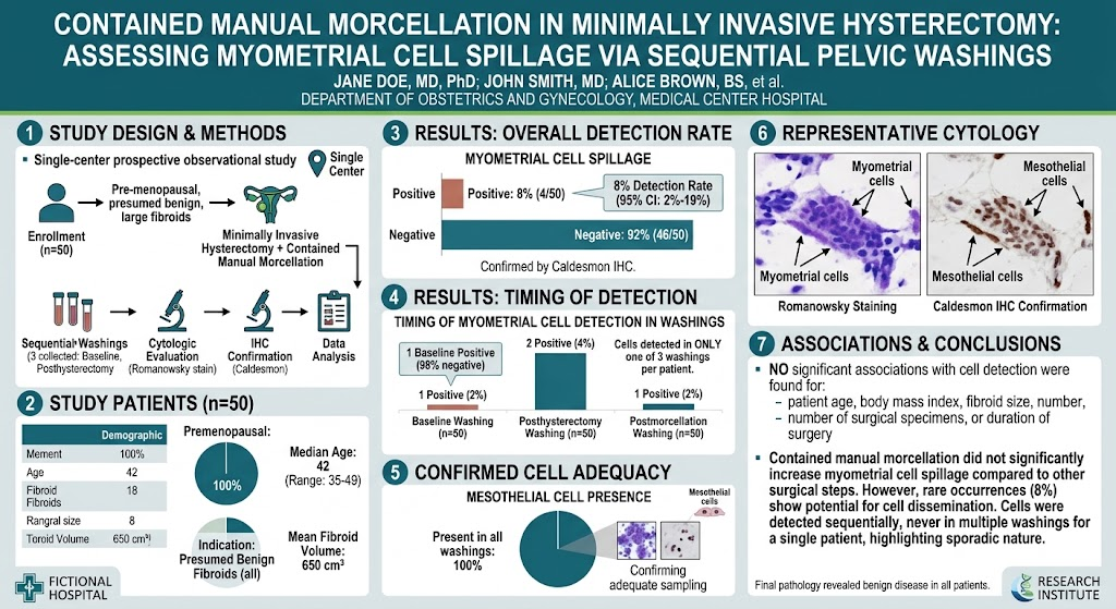
1. Contained manual morcellation in minimally invasive hysterectomy shows a low rate (8%) of myometrial cell spillage.
2. Most myometrial cell spillage occurs before morcellation starts, immediately following hysterectomy.
3. Sequential pelvic washings with immunohistochemical confirmation provide robust evidence of cell dissemination timing.
4. These findings support the safety and effectiveness of containment systems in preventing tissue dissemination during morcellation.
Study Background
Minimally invasive hysterectomy (MIH) is a widely adopted surgical approach for benign uterine conditions, such as large fibroids, due to benefits including reduced postoperative pain, faster recovery, and shorter hospital stays. However, the need to remove large tissue specimens through small incisions has led to the use of morcellation techniques, which mechanically fragment the uterus to facilitate extraction.
Concerns have arisen regarding the potential for tissue dissemination during morcellation, especially malignant cells inadvertently spread in cases of unsuspected malignancy, or even benign myometrial cells that may implant ectopically. This risk prompted warnings from regulatory bodies and fostered the development of containment systems to minimize cell spillage during manual morcellation within a sealed environment.
Despite this, the precise timing and extent of myometrial cell spillage during contained manual morcellation remain unclear. Particularly, it is unknown whether tissue dissemination occurs mainly during morcellation or if earlier surgical steps contribute. Clarifying this distinction is critical to assessing the true safety benefit of containment techniques.
Study Design
This prospective single-center observational study enrolled 50 premenopausal patients undergoing minimally invasive hysterectomy for presumed benign large uterine fibroids requiring manual morcellation within a containment system. Inclusion criteria focused on patients without suspicion of malignancy, to isolate the study to benign conditions relevant to most clinical practice.
The intervention involved contained sharp manual morcellation of the uterus within a specialized bag designed to prevent tissue fragments from dispersing into the peritoneal cavity. Three sequential pelvic washings were collected at defined surgical time points: (1) baseline immediately after peritoneal entry, (2) posthysterectomy before morcellation commenced, and (3) postmorcellation. The sequential sampling allowed temporal mapping of cell spillage.
To detect myometrial cells, cytologic evaluation was performed using Romanowsky staining, a technique highlighting cellular morphology. Confirmatory immunohistochemical staining with Caldesmon, a protein marker characteristic of smooth muscle (myometrial) cells, was employed. Pathologists were blinded to clinical data to minimize bias. The primary outcome was presence of myometrial cells; secondary outcomes included timing of detection and correlations to clinical variables such as uterine size or operative time.
Key Findings
Myometrial cells were identified in 4 out of 50 patients (8%, 95% confidence interval: 2%–19%), indicating a generally low incidence of cell spillage during the surgical process with containment. Importantly, myometrial cells were detected only once per patient within the three sequential washings, illustrating spillage was transient or limited in timing.
Baseline washings—obtained immediately after entering the peritoneal cavity—were negative in 98% of patients, confirming no preexisting contamination and adequate sampling technique. Detection varied in the remaining samples: 4% posthysterectomy before morcellation and 2% postmorcellation. This pattern suggests that cell spillage predominantly occurs after uterine removal but before morcellation, implicating surgical maneuvers during hysterectomy rather than the morcellation process itself as the main source.
All positive findings were validated by Caldesmon immunostaining to distinguish true myometrial cells from mesothelial or inflammatory cells. Mesothelial cells were observed in all washings, confirming specimen adequacy and appropriate sampling of the peritoneal environment.
Final pathology confirmed benign disease in all cases, which aligns with the study focus on nonmalignant indications and supports that detected cells originated from fibroid or normal myometrium.
Expert Commentary
This study offers rigorous evidence addressing a critical knowledge gap in hysterectomy safety. The use of sequential pelvic washings coupled with robust cytologic and immunohistochemical methods provides unique temporal resolution of tissue spillage events.
Notably, the data challenge the common assumption that morcellation is the primary driver of myometrial cell dissemination. Instead, they highlight that tissue manipulation during hysterectomy may account for most spillage. This finding is clinically relevant because containment bags act only during morcellation and do not affect earlier surgical steps.
These insights underscore the importance of meticulous surgical technique throughout hysterectomy and suggest containment systems contribute significantly to preventing additional spillage once morcellation starts, but cannot entirely eliminate cell release occurring beforehand.
Limitations include the single-center design and relatively small sample size, which may affect generalizability. Additionally, the study excluded patients with suspected malignancy, so implications for cancer risk remain unknown. Future multicenter trials and extended follow-up to assess clinical outcomes such as parasitic leiomyomas or peritoneal implants would further solidify the protective value of containment strategies.
Conclusion
This investigation into contained manual morcellation during minimally invasive hysterectomy demonstrates a low incidence of myometrial cell spillage, mostly occurring before morcellation. Sequential pelvic washings confirm that containment systems effectively reduce dissemination risk during the morcellation phase.
These findings reinforce the clinical safety of contained manual morcellation for benign fibroid uteri and emphasize that surgeons should be cognizant of potential cell release during earlier operative steps. The study supports continued use and refinement of containment devices as an important safeguard in minimally invasive gynecologic surgery.
Further research should aim to expand patient populations, explore malignancy contexts, and evaluate long-term clinical impacts to optimize surgical protocols and improve patient safety.
Funding and ClinicalTrials.gov
The study was supported by institutional research funds at the tertiary care center where the investigation took place. Registration information and funding disclosures were not detailed in the abstract.
References
1. Eyada MF, Clement CG, Segura C, et al. Does contained manual morcellation in minimally invasive hysterectomy prevent myometrial cell spillage? Evidence from sequential pelvic washings. Am J Obstet Gynecol. 2026 Mar 26;235(2):322-329. PMID: 41903875.
2. Cohen SL, Kho RM, Tayag C, et al. Geo-containment or no containment during power morcellation: an evidence-based systematic review of peritoneal tissue dissemination. J Minim Invasive Gynecol. 2021;28(4):595-605.
3. FDA Safety Communication, 2014. Update on the risk of power morcellation in hysterectomy and myomectomy for uterine fibroids.
4. Kho RM, Mandavilli SR. Current approaches to contained morcellation in gynecologic surgery. Obstet Gynecol. 2019;133(4):754-761.