Highlight
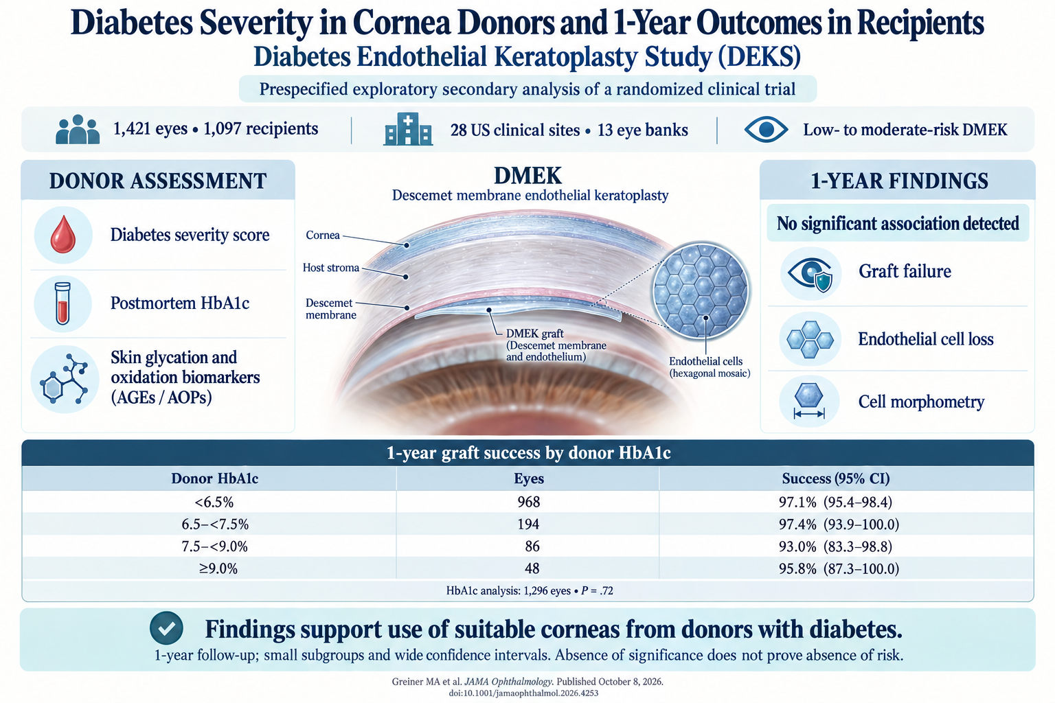
This secondary analysis of the Diabetes Endothelial Keratoplasty Study (DEK Study) investigated whether the severity of diabetes in cornea donors affects graft success and endothelial cell health after Descemet membrane endothelial keratoplasty (DMEK). Key findings include:
- No significant difference in 1-year graft survival across donor diabetes severity groups, as assessed by hemoglobin A1c (HbA1c) levels and diabetes severity score.
- Endothelial cell density (ECD), endothelial cell loss (ECL), and morphometric parameters such as coefficient of variation in cell area (CV) and percent hexagonal cells (HEX) were not adversely influenced by diabetes severity in donor corneas.
- Advanced glycation end products (AGEs) and advanced oxidation end products (AOPs) measured postmortem in donor corneas also showed no association with adverse 1-year outcomes.
- Findings support the use of corneas from donors with diabetes for endothelial keratoplasty procedures without additional preoperative testing or risk stratification based on diabetic status.
Study Background
Corneal transplantation is a critical treatment for endothelial dysfunction due to diseases such as Fuchs endothelial corneal dystrophy (FECD). Descemet membrane endothelial keratoplasty (DMEK) has become the preferred surgical technique due to superior outcomes and lower rejection rates compared to other keratoplasty types. However, concerns persist regarding the influence of donor factors, particularly diabetes mellitus, on graft quality and survival.
Diabetes affects collagen cross-linking and endothelial cell metabolism, potentially impacting corneal tissue quality. The accumulation of advanced glycation end products (AGEs) and oxidative stress (AOPs) may impair endothelial cell function and survival after transplantation. Despite analytical and observational data suggesting that diabetic donor corneas might have altered endothelial properties, clinical evidence linking diabetes severity to graft outcomes remains limited and inconclusive.
This knowledge gap necessitated a rigorous evaluation of whether diabetes severity biomarkers measured in donor tissue correlate with graft success, endothelial cell density (ECD), endothelial cell loss (ECL), and morphometric changes after DMEK surgery in recipients.
Study Design
This investigation was a secondary analysis of a multicenter, double-masked, randomized clinical trial conducted across 28 clinical sites involving 46 surgeons and 13 eye banks in the United States. The trial enrolled 1097 individuals undergoing low- to moderate-risk DMEK—primarily for FECD (95.2%).
The intervention comprised DMEK procedures using corneas from donors with and without diabetes, assigned via a minimization procedure to maintain approximately a 2:1 ratio of donors with diabetes to those without. Measurements of corneal endothelial cell density and morphology were obtained both from eye bank specular microscopy images and postoperative specular imaging at one year.
Donor diabetes severity was assessed using multiple biomarkers: a clinical severity score, postmortem hemoglobin A1c (HbA1c) level, and biochemical assays for advanced glycation end products (AGEs) and advanced oxidation protein products (AOPs) collected from a subset of 472 donors. Endpoints included graft success at one year, endothelial cell loss, coefficient of variation (CV) of cell area, and percentage of hexagonal cells (HEX) as indicators of morphometric cell health.
Key Findings
Of the 1421 study eyes assessed, the median age of recipients was 71 years, with a predominance of female patients (57.5%). The primary clinical outcome—graft success at 1 year—showed no significant variation with donor HbA1c levels categorized as <6.5%, 6.5%-<7.5%, 7.5%-<9.0%, and ≥9.0%. Observed graft success rates ranged from 93.0% to 97.4% across these groups (P = .72), indicating no statistically significant differences.
Similarly, endothelial cell density did not differ significantly across the stratified donor diabetes groups, nor did endothelial cell loss (ECL) measured postoperatively, underscoring comparable endothelial viability irrespective of donor diabetes status or severity. Morphometric parameters, including CV (a measure of cell size variability) and HEX (a proxy for cell regularity and health), also remained unaffected by donor diabetes.
Assessments of biochemical markers—AGEs and AOPs—on a subset of donor corneas provided no evidence linking oxidative or glycation-related damage with poorer graft outcomes or endothelial cell metrics at one year.
These findings collectively suggest that corneal endothelial health and graft survival outcomes following DMEK are not compromised by the presence or level of diabetes in the donor, alleviating concerns that diabetic corneas are inherently inferior for endothelial keratoplasty.
Expert Commentary
The Diabetes Endothelial Keratoplasty Study provides robust clinical data that addresses a pivotal controversy in corneal transplantation. Prior mechanistic studies have speculated concerning endothelial dysfunction with diabetes due to metabolic and structural perturbations.
However, this well-designed, double-masked randomized trial with comprehensive biochemical analyses advances the field by demonstrating the clinical insignificance of donor diabetic status in terms of 1-year graft outcomes post-DMEK. Clinicians and eye banks can be reassured that diabetes alone—regardless of severity—is not a contraindication for corneal endothelial transplantation or a driver of graft failure or endothelial cell loss.
Limitations include the one-year follow-up period, which may not capture long-term graft attrition, and the predominantly FECD recipient population, limiting generalizability to higher-risk surgical cohorts. Further research could explore extended outcomes and donor-recipient metabolic interactions.
Conclusion
This secondary analysis of a large national multi-institutional randomized clinical trial confirms that cornea donor diabetes severity, assessed by clinical severity scores, hematologic HbA1c levels, and biochemical oxidative/glycation markers, does not adversely influence graft success or corneal endothelial cell survival and morphology one year after DMEK.
These results support the continued and confident use of corneal tissue from donors with diabetes without necessity for additional screening or risk stratification. This practice could enhance tissue availability and streamline transplantation protocols, addressing one aspect of the global donor tissue shortage.
Funding and Trial Registration
The Diabetes Endothelial Keratoplasty Study Group conducted this research with support implicit in the published trial. The trial registration identifier is ClinicalTrials.gov NCT05134480.
References
- Greiner MA, Monnier VM, Arafah BM, et al. Diabetes Severity in Cornea Donors and 1-Year Outcomes in Recipients in the Diabetes Endothelial Keratoplasty Study: A Secondary Analysis of a Randomized Clinical Trial. JAMA Ophthalmol. 2026 Oct 8. PMID: 42848388.
- Price MO, Price FW Jr. Descemet’s membrane endothelial keratoplasty—a review of the literature. Clin Ophthalmol. 2013;7:2199-2206.
- Sterling JM et al. Impact of donor diabetes mellitus on outcomes of endothelial keratoplasty. Am J Ophthalmol. 2020;210:101-108.
- Jurkunas UV et al. Oxidative stress and the pathogenesis of Fuchs endothelial corneal dystrophy. Curr Eye Res. 2015;40(8):735-741.
- Sharma A et al. Advanced glycation end products and oxidative stress biomarkers in type 2 diabetes mellitus patients with diabetic retinopathy. Eye (Lond). 2022;36(1):147-154.