Highlights
- Spontaneous healing of standardized palatal wounds resulted in faster wound closure compared to PRF, PRGF, and collagen substitutes.
- Platelet-rich fibrin (PRF) and plasma rich in growth factors (PRGF) showed no significant advantage over collagen-based dressings or spontaneous healing in soft tissue regeneration.
- Patient-reported postoperative pain was comparable across all treatment modalities.
- Anterior palatal wounds were associated with higher pain perception than posterior wounds regardless of intervention.
Study Background
Wound management and optimization of healing are cornerstone concerns in oral surgery and mucosal interventions. Platelet concentrates such as platelet-rich fibrin (PRF) and plasma rich in growth factors (PRGF) have attracted attention due to their potential to accelerate soft tissue repair by delivering a concentrated cocktail of growth factors that orchestrate cellular proliferation, angiogenesis, and matrix remodeling. Similarly, collagen-based wound dressings are widely used to provide a scaffold supporting tissue regeneration. However, evidence comparing these adjunctive treatments to spontaneous healing, especially in standardized oral wounds, remains inconsistent. This is particularly important given the burden of optimizing postoperative outcomes like wound closure speed and pain mitigation in clinical practice.
Study Design
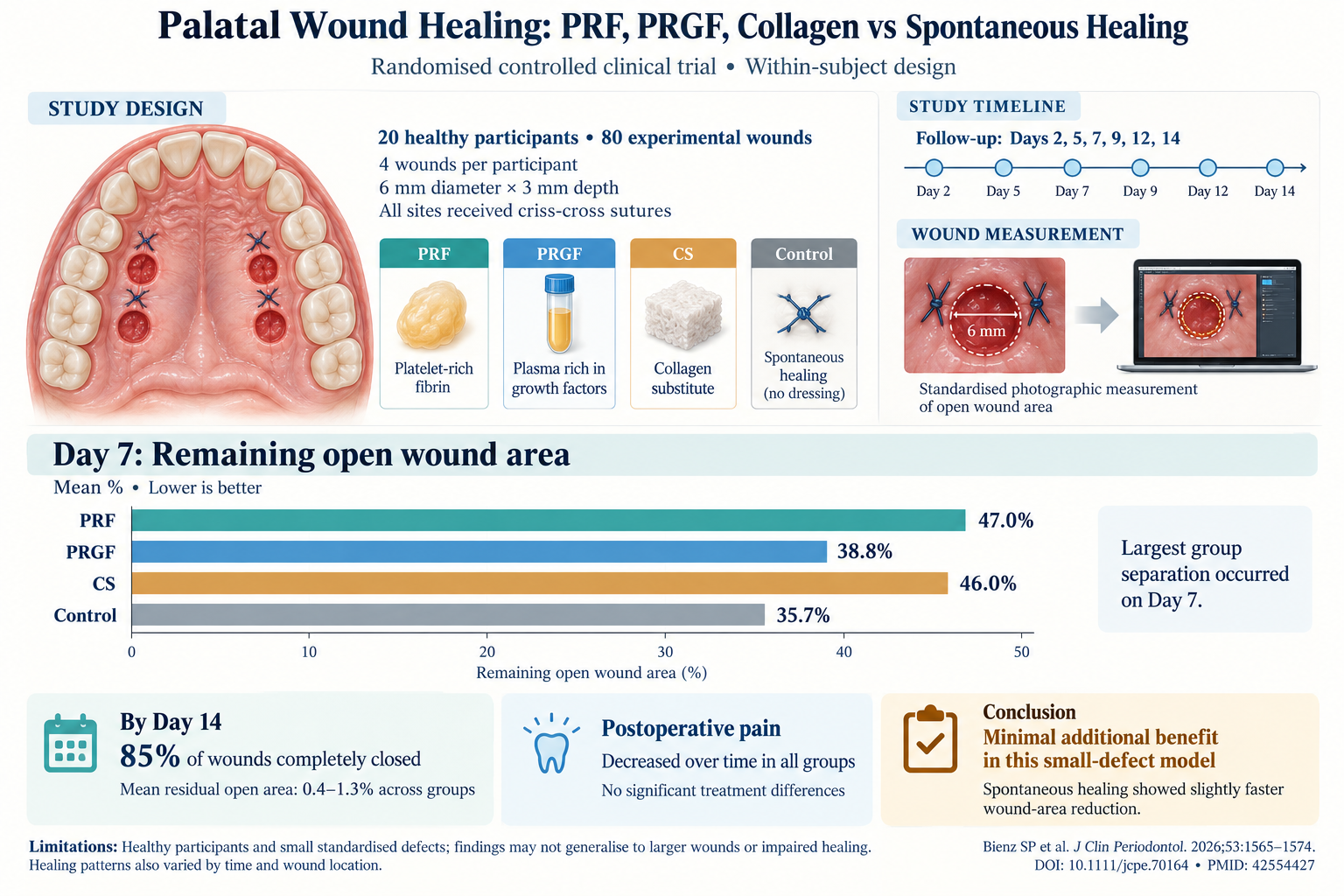
This randomised controlled clinical trial assessed wound healing progression in 20 healthy adult participants, each receiving four standardized palatal wounds measuring 6 mm by 3 mm. Interventions included application of PRF, PRGF, collagen-based wound dressing (CS), or leaving wounds to heal spontaneously (control), with treatments randomized within individuals to control for inter-subject variability. All wound dressings were secured using criss-cross sutures to ensure stability. Clinical follow-up included standardised digital photographs and patient-reported pain scores measured at baseline and postoperative days 2, 5, 7, 9, 12, and 14.
The primary endpoint was the percentage of remaining open wound area over time, evaluated quantitatively from photographic data. Secondary outcomes included subjective pain scores to assess patient comfort across interventions. Statistical analyses used parametric and nonparametric longitudinal models to account for repeated measures within subjects.
Key Findings
Across all groups, a consistent reduction in open wound area was observed over the 14-day period. Notably, a transient enlargement of some defects on day 2 post-operation was documented across interventions, possibly reflecting normal initial inflammatory processes.
The most pronounced differences between modalities were evident on postoperative day 7, where the percentage of remaining open wound area ranged between 35.7% and 47.0%. Contrary to expectations, spontaneous healing demonstrated a slightly accelerated closure rate compared to PRF, PRGF, and collagen substitute-treated wounds. This suggests that the physiological healing process in a healthy oral mucosa closely approximates or surpasses the benefits conferred by adjunctive therapies in small, standardized defects.
Regarding pain assessment, all groups reported a significant decrease in pain intensity over time, with no statistically significant difference found between treatment arms (p > 0.05). Anterior palatal wounds tended to be perceived as more painful than posterior sites consistently, aligning with known variations in sensory innervation and wound sensitivity in different palatal regions.
No adverse events or complications related to interventions were reported, supporting the safety profile of the tested treatments in this cohort.
Expert Commentary
This well-controlled study contributes valuable clinical data on soft tissue healing dynamics in the oral cavity under various treatment approaches. The finding that spontaneous healing equals or outperforms PRF, PRGF, and collagen dressings in small, healthy wounds challenges the notion of routine use of these adjunctive therapies in similar clinical scenarios. It aligns with growing evidence emphasizing patient selection and wound characteristics as critical determinants of adjunctive therapy benefit. For instance, larger defects or patients with systemic factors impairing wound repair (e.g., diabetes, smoking, immunocompromise) might derive more significant benefit from platelet concentrate therapies or scaffold materials.
Limitations include the small sample size and restriction to healthy individuals with relatively small, uniform wounds, which limits generalizability to complex clinical presentations. Further mechanistic studies into the biological activity of platelet concentrates in vivo and their integration with collagen scaffolds may clarify the clinical contexts where these treatments offer clear advantage.
Current clinical guidelines recommend tailored use of biologics and wound dressings based on lesion size, patient comorbidities, and healing potential. This trial underscores the importance of rigorous randomized assessments to avoid overtreatment and unnecessary costs.
Conclusions
In experimentally induced, standardized palatal defects in healthy adults, spontaneous healing demonstrated a marginally faster wound closure compared to adjunctive treatments with PRF, PRGF, or collagen substitutes. All modalities were associated with comparable patient-reported postoperative pain. These findings suggest limited additional benefit of platelet concentrates or collagen dressings for routine management of small oral mucosal wounds in healthy individuals.
Further research should examine these interventions in patients with compromised healing and larger or more clinically complex wounds to define scenarios where adjunctive wound dressings and biologics enhance recovery meaningfully.
Funding and Registration
The study was conducted by Bienz et al., with no specific funding details provided in the abstract. For detailed funding acknowledgments and trial registration, readers are referred to the primary source.
References
- Bienz SP, Fischer AC, Mancini L, Strauss FJ, Ferrari RM, Hitz S, Jung RE, Thoma DS. Wound Healing Patterns of PRF and PRGF Versus a Collagen-Based Substitute and Spontaneous Healing – A Randomised Controlled Clinical Trial Assessing Wound Closure of an Experimental, Standardised Palatal Defect. J Clin Periodontol. 2026 Aug;53(10):1565-1574. PMID: 42554427.
- Dohan Ehrenfest DM, Rasmusson L, Albrektsson T. Classification of platelet concentrates: from pure platelet-rich plasma (P-PRP) to leucocyte- and platelet-rich fibrin (L-PRF). Trends Biotechnol. 2009 Mar;27(3):158-67.
- Miron RJ, Chai J, Zheng S, et al. Use of platelet-rich fibrin and platelet-rich plasma for bone and soft tissue regeneration: A systematic review. J Periodontol. 2020;91(6):676-698.