Highlight
This post hoc analysis of the Singapore Asymptomatic Narrow Angles-Laser Iridotomy Study (ANA-LIS) reveals that higher baseline intraocular pressure (IOP), narrower anterior chamber angles, and the presence of plateau iris configuration in multiple quadrants are significant risk factors for progression from primary angle closure suspect (PACS) to primary angle closure (PAC) over a five-year period. Comprehensive anterior segment imaging techniques such as AS-OCT and ultrasound biomicroscopy (UBM) are instrumental in risk stratification and patient management.
Study Background
Primary angle closure glaucoma (PACG) remains a leading cause of irreversible blindness worldwide, especially in Asian populations. It develops through a pathophysiological sequence beginning with primary angle closure suspect (PACS), characterized by anatomically narrow angles but without elevated intraocular pressure (IOP) or peripheral anterior synechiae, potentially progressing to primary angle closure (PAC) marked by raised IOP or angle closure-related structural changes. Accurate identification of PACS eyes at greatest risk of progression is crucial for timely intervention, such as laser peripheral iridotomy (LPI), to prevent irreversible optic nerve damage and visual loss. However, the factors predictive of disease progression have remained insufficiently characterized, particularly using advanced anterior segment imaging modalities.
Study Design
This study is a post hoc analysis of the ANA-LIS, a randomized clinical trial conducted between 2004 and 2018 across five tertiary eye centers in Singapore. The original trial enrolled 480 participants with PACS, who underwent comprehensive ophthalmic evaluation. Baseline assessments included anterior segment optical coherence tomography (AS-OCT) and ultrasound biomicroscopy (UBM) to quantify angle anatomy and detect plateau iris configuration. One eye per participant was randomly selected for laser peripheral iridotomy (LPI), with the fellow eye serving as control. Progression was defined as development of primary angle closure—manifested by elevated IOP greater than 24 mm Hg, formation of peripheral anterior synechiae (PAS) occupying at least one clock hour, or an acute angle closure episode. Data analysis was conducted from May to July 2025 using generalized linear models and Cox regression models clustered by participant.
Key Findings
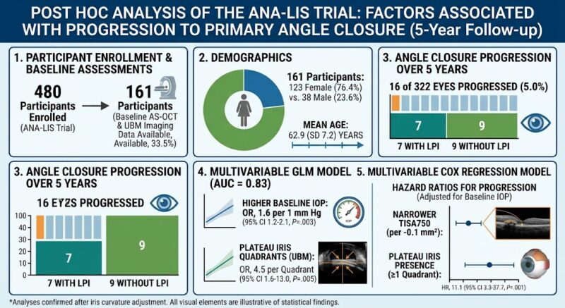
Out of 480 participants, 161 underwent baseline AS-OCT and UBM examinations and were included in the current analysis. The cohort was predominantly female (76.4%) with a mean age of 62.9 years. During the 5-year follow-up, 16 of 322 eyes (5.0%) progressed from PACS to PAC. The progression rate was comparable between eyes that received LPI (7 eyes) and those that did not (9 eyes).
Multivariable analysis revealed several independent predictors of progression:
- Baseline Intraocular Pressure (IOP): Each 1 mm Hg increase in baseline IOP was associated with a 60% increased odds of progression (OR 1.6; 95% CI, 1.2-2.1; P = .003), underscoring the prognostic importance of subtle IOP elevations.
- Plateau Iris Configuration: Identified by UBM, the presence of plateau iris in multiple quadrants was associated with markedly increased risk. Each additional quadrant with plateau iris raised odds of progression by 4.5-fold (OR 4.5; 95% CI, 1.6-13.0; P = .005). In time-to-event analysis, eyes with plateau iris in at least one quadrant had an 11-fold higher hazard of progression (HR 11.1; 95% CI, 3.3-37.7; P = .001).
- Anterior Chamber Angle Narrowing: Trabecular-iris space area measured at 750 µm from the scleral spur (TISA750) was inversely related to progression risk. Smaller TISA750 (i.e., narrower angles) were associated with a threefold increase in hazard per 0.1 mm2 decrement (HR 3.1; 95% CI 1.3-7.5; P = .02).
Adjusting for iris curvature did not materially change these associations, affirming the robustness of these anatomical predictors. The model incorporating these factors achieved excellent discriminatory ability with an area under the receiver operating characteristic curve of 0.83.
Expert Commentary
The findings from this analysis emphasize the value of advanced anterior segment imaging modalities in risk stratifying PACS patients. While the role of LPI in preventing progression has been debated, these data highlight that anatomical parameters and subtle IOP elevations should guide clinical decision-making rather than solely relying on gonioscopic angle appearance. Plateau iris, often under-recognized in clinical practice, emerges as a strong indicator of progression risk, particularly when present in multiple quadrants, suggesting that eyes with this configuration might benefit from closer monitoring or alternative interventions such as iridoplasty.
Limitations include the post hoc nature of the analysis and the relatively small number of progression events, which may limit generalizability. Nevertheless, the longitudinal design, rigorous imaging techniques, and multicenter participation strengthen the validity of conclusions.
Conclusion
This post hoc analysis from the ANA-LIS trial demonstrates that higher baseline IOP, narrower anterior chamber angles quantified by TISA750, and presence of plateau iris configuration in multiple quadrants independently increase the risk of progression from PACS to PAC over five years. Incorporating comprehensive anterior segment imaging into routine assessment can improve early identification of high-risk eyes, enabling tailored preventive strategies to curb vision loss from angle closure disease.
Funding and Trial Registration
The original ANA-LIS trial was supported by research grants from multiple ophthalmic research bodies. The trial is registered at ClinicalTrials.gov under identifier NCT00347178.
References
- Tun TA, Yoo C, Servillo A, et al. Factors Associated With Progression to Primary Angle Closure: A Post Hoc Analysis of the ANA-LIS Trial. JAMA Ophthalmol. 2026;doi:10.1001/jamaophthalmol.2026.61625.
- Qin L, Wang D, He M, et al. Anterior segment OCT and Ultrasound Biomicroscopy in angle-closure glaucoma. Am J Ophthalmol. 2021;223:13-23.
- Foster PJ, Buhrmann R, Quigley HA, et al. The definition and classification of glaucoma in prevalence surveys. Br J Ophthalmol. 2002;86(2):238-242.