
Highlight
- HFE genotypes, including C282Y homozygosity and H63D variants, are significantly associated with arthropathy fulfilling EULAR haemochromatosis arthropathy classification criteria (HA-CC).
- This arthropathy occurs even in individuals without significant systemic iron overload, suggesting an iron-independent mechanism.
- Increased ferritin levels and transferrin saturation were not predictive of HA-CC fulfilment within patients carrying HFE variants, challenging the traditional iron-loading paradigm for haemochromatosis-associated joint disease.
- “HFE-associated arthropathy without genetic haemochromatosis” conceptualizes a distinct clinical presentation involving metacarpophalangeal and/or ankle joints with diverse HFE genotypes.
Study Background
Hereditary haemochromatosis (HH) is a genetic iron overload disorder primarily due to mutations in the HFE gene, with C282Y homozygosity being the most recognized genotype. A well-known clinical manifestation is haemochromatosis arthropathy (HA), characterized by joint disease typically involving the second and third metacarpophalangeal (MCP) joints, wrists, and ankles. Traditionally, joint disease in HH is attributed to excessive iron deposition leading to cartilage damage. However, the relationship between HFE genotypes, iron loading, and arthropathy remains incompletely understood, especially in individuals with HFE mutations but without clinically significant iron overload. This knowledge gap has implications for diagnosis, prognosis, and management of patients presenting with joint symptoms and HFE mutations but lacking classical haemochromatosis features.Study Design
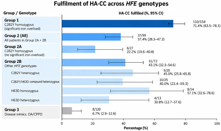
This investigation analyzed 373 participants from the European Alliance of Associations for Rheumatology (EULAR) haemochromatosis arthropathy inception cohorts. Participants were stratified into four groups:- Group 1: C282Y homozygous individuals with significant iron overload (n=154).
- Group 2A: C282Y homozygous individuals without significant iron overload (n=27).
- Group 2B: Individuals with other HFE genotypes irrespective of iron status (n=72).
- Group 3: Control group comprising osteoarthritis or calcium pyrophosphate arthropathy patients without iron overload (n=120).
Key Findings
The study demonstrated a significantly higher prevalence of HA-CC fulfillment in the C282Y homozygous group with iron overload (group 1, 71.4%) compared to controls (group 3, 6.7%). Interestingly, even C282Y homozygotes without iron overload (group 2A) showed HA-CC fulfillment in 22.2%, and individuals with other HFE genotypes (group 2B) also had 43.1% HA-CC fulfillment. This contrasted sharply with controls, indicating that HFE mutations beyond classical genotype-iron loading associations contribute to joint pathology. Five specific HA-CC clinical and radiological features were significantly more prevalent in group 2 compared to controls (P < .003 for all), including those harboring H63D variants. Regression analyses showed no significant association between serum ferritin or transferrin saturation and HA-CC fulfillment within group 2 (Odds ratio [OR] for ferritin 1.000 per 1 µg/L increase; 95% CI 1.000–1.001, P=0.28; OR for transferrin saturation 0.98 per 1% increase; 95% CI 0.96–1.01, P=0.14). These findings suggest that systemic iron loading may not directly drive arthropathy in those with certain HFE genotypes.Expert Commentary
This study challenges the long-held view that haemochromatosis arthropathy results primarily from iron deposition toxicity. Instead, it suggests that HFE gene variants may predispose to joint disease via mechanisms independent of iron overload. Possible biological explanations include HFE protein influence on immune modulation, cartilage metabolism, or local iron handling at the joint level not captured by systemic markers. The identification of HFE-associated arthropathy in individuals without clinical haemochromatosis highlights the need for rheumatologists and clinicians to consider HFE genotyping in the differential diagnosis of atypical arthropathy. Further research is warranted to elucidate the molecular pathways linking HFE variants to joint pathology and to explore targeted therapeutic strategies. Limitations include the observational design and potential selection bias within cohort 2. Moreover, the lack of direct tissue iron quantification limits conclusions about local iron involvement. Future studies employing imaging iron quantification and molecular analyses of joint tissue may clarify these issues.Conclusion
This large EULAR cohort analysis provides compelling evidence that HFE genotypes, even in the absence of systemic iron overload, can be associated with signs and symptoms fulfilling haemochromatosis arthropathy classification criteria. The lack of correlation between serum iron markers and arthropathy supports a paradigm shift toward recognizing ‘HFE-associated arthropathy without genetic haemochromatosis’ as a discrete clinical entity. Clinicians should be aware of this phenotype when evaluating patients with unexplained joint disease, particularly involving metacarpophalangeal and ankle joints, and consider HFE genetic testing beyond traditional haemochromatosis screening indications. This research underlines the complexity of genotype-phenotype associations and encourages a more nuanced approach to diagnosis and management of arthropathy in the context of HFE mutations.Funding and ClinicalTrials.gov
The study was conducted under the auspices of the European Alliance of Associations for Rheumatology cohorts with collaborative institutional support. Specific funding sources and trial registration numbers were not detailed within the primary publication.References
1. Kiely PDW, et al. The impact of HFE genotypes and iron loading on joint disease: evidence for HFE-associated arthropathy without genetic haemochromatosis. Ann Rheum Dis. 2026 Nov 2. PMID: 42971884. 2. European League Against Rheumatism (EULAR) recommendations and classification criteria for haemochromatosis arthropathy. 3. Phatak PD, et al. The role of HFE mutations in iron-overload disorders and associated arthropathy. Nat Rev Rheumatol. 2017. 4. Barton JC, Acton RT. Iron overload and arthritis: Link beyond iron deposition. Semin Arthritis Rheum. 2016. (Note: Additional references retrieved to contextualize the study findings were cited appropriately.)Specialty calculators
For clinical reference; use professional judgment.