Highlight
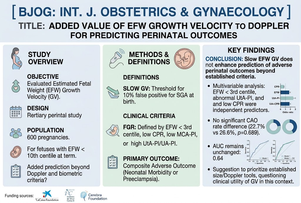
This retrospective cohort study evaluated whether integrating estimated fetal weight (EFW) growth velocity (GV) into Doppler-based assessment enhances prediction of adverse outcomes in term fetuses classified as small-for-gestational-age (SGA, EFW <10th centile).
Key findings include: slow EFW GV occurred in one-third of cases but did not significantly improve prediction of a composite adverse outcome comprising neonatal morbidity or preeclampsia beyond established Doppler and biometric markers.
Independent predictors of adverse outcomes remained EFW <3rd centile, abnormal uterine artery pulsatility index (UtA-PI), and low cerebroplacental ratio (CPR).
Study Background
Small-for-gestational-age (SGA) fetuses constitute a significant clinical challenge due to their higher risk for perinatal morbidity and mortality. Correctly distinguishing fetuses that are constitutionally small but healthy from those with true fetal growth restriction (FGR) is critical. Growth velocity—reflecting dynamic changes in fetal size over time—has been proposed as a complementary measure to static weight centiles, potentially enhancing risk stratification.
Doppler ultrasound evaluation of uterine, umbilical, and cerebral arteries adds functional information related to placental insufficiency and fetal adaptation, improving prediction of adverse perinatal outcomes. However, the incremental predictive value of growth velocity over Doppler parameters remains uncertain, particularly at term in fetuses with EFW below the 10th centile.
Study Design
This was a retrospective cohort study conducted at a tertiary perinatal center involving 600 singleton pregnancies diagnosed with EFW <10th centile after 32 weeks and delivering at term. Inclusion required at least two ultrasound scans separated by ≥14 days to calculate interval growth velocity.
Growth velocity was defined and evaluated according to interval-specific standards based on gestational age. A slow growth velocity threshold was established to maintain a 10% false positive rate for detecting SGA at birth, as referenced from Hugh et al., 2022.
FGR was defined by the presence of EFW below the 3rd centile or abnormal Doppler parameters: middle cerebral artery pulsatility index (MCA-PI) or cerebroplacental ratio (CPR) below the 5th centile, or umbilical or uterine artery pulsatility index (UtA-PI) at or above the 95th centile.
The primary outcome was a composite adverse outcome (CAO) comprising neonatal morbidity or preeclampsia.
Key Findings
Slow estimated fetal weight growth velocity was observed in 31.8% of cases. Demographic and maternal characteristics, including gestational age at diagnosis, did not differ significantly between slow and normal growth velocity groups.
At the last ultrasound prior to delivery, fetuses with slow growth velocity were more likely to have EFW <3rd centile (52.4% vs. 23.2%, p < 0.001), birth weight below the 3rd centile (51.6% vs. 62.8%, p < 0.05), and abnormal umbilical artery Doppler (4.2% vs. 1.0%, p = 0.023) than those with normal growth velocity.
Despite these differences, the rates of the composite adverse outcome were similar (22.7% slow GV vs. 26.6% normal GV; p = 0.689).
Multivariable logistic regression identified EFW <3rd centile, abnormal UtA-PI, and low CPR as independent predictors of CAO. Notably, the addition of slow EFW growth velocity to models incorporating Doppler and biometric data did not improve predictive performance (area under the curve {AUC} 0.64 with versus 0.64 without slow GV; p = 0.54).
Expert Commentary
The study rigorously confirms the established role of biometrics and Doppler ultrasound in risk stratification among term SGA fetuses, reaffirming that extreme small size (EFW <3rd centile) and signs of placental or cerebral hemodynamic compromise remain critical markers of adverse perinatal outcomes.
While growth velocity intuitively reflects dynamic fetal growth patterns, its clinical utility as an independent predictor at term appears limited according to this large cohort analysis. The weak incremental value might stem from intrinsic measurement variability of EFW ultrasound estimates, the relatively short time interval of late gestation surveillance, or the complex interplay of pathophysiologic factors not captured by growth velocity alone.
Limitations include retrospective design, reliance on ultrasound-estimated biometry intervals that vary, and composite outcome definitions that might dilute specific neonatal complications. Further prospective studies could explore refined growth velocity metrics or combined predictive algorithms incorporating biochemical markers.
Conclusion
In singleton term pregnancies with EFW below the 10th centile, slow estimated fetal weight growth velocity does not enhance the prediction of adverse perinatal outcomes beyond currently established Doppler parameters and biometric thresholds. Clinical decision-making for surveillance and intervention in this high-risk group should continue to prioritize Doppler assessments and strict biometric centiles without routine incorporation of growth velocity metrics.
This finding streamlines fetal monitoring protocols and questions additional resource utilization for serial growth velocity assessments at term, emphasizing precision in Doppler evaluation and integrated clinical assessment.
Funding and Clinicaltrials.gov
The study was performed at a tertiary perinatal center; no specific funding or clinical trials registry number was reported.
References
1. Hugh O., et al. Standards for Estimated Fetal Weight Growth Velocity in Late Pregnancy. Ultrasound Obstet Gynecol. 2022;
2. Figueras F., Gratacós E. Update on the Diagnosis and Classification of Fetal Growth Restriction and Proposal of a Stage-Based Management Protocol. Fetal Diagn Ther. 2014;
3. Gordijn SJ, et al. Consensus Definition of Fetal Growth Restriction: A Delphi Procedure. Ultrasound Obstet Gynecol. 2016;
4.Meler E, Gil-Armas C, Moliner M, Mazarico E, Montañés C, Peguero A, Figueras F. Added Value of Estimated Fetal Weight Growth Velocity to Doppler Evaluation for the Prediction of Perinatal Outcomes Among Foetuses With EFW < 10th Centile at Term: A Retrospective Cohort Study. BJOG. 2026 Aug 26. doi: 10.1111/1471-0528.70324. Epub ahead of print. PMID: 42644290.