Highlight
– Indocyanine green (ICG) fluorescence imaging enhances detection and intraoperative margin assessment of colorectal liver metastases during minimally invasive wedge resections.
– The study identified fluorescence rim integrity as a reliable real-time marker predicting R0 resection.
– ICG fluorescence enabled detection of additional occult subcentimeter lesions not identified by conventional imaging.
– Parenchymal-sparing resections aided by ICG fluorescence may optimize oncologic clearance and preserve healthy liver tissue.
Study Background
Colorectal cancer frequently metastasizes to the liver, with surgical resection remaining the potentially curative treatment. Over recent years, there has been a move toward parenchymal-sparing liver resections aiming to preserve maximal functional hepatic tissue, which is crucial for postoperative liver function and repeat resections if needed. However, these limited resections can increase the risk of positive surgical margins (R1), which are associated with worse oncologic outcomes.
Indocyanine green (ICG) fluorescence imaging has emerged as a promising intraoperative adjunct to enhance visualization of hepatic tumors. Administered intravenously before surgery, ICG accumulates differentially in tumor tissue or adjacent liver parenchyma, producing distinct fluorescence patterns under near-infrared light. This technique holds potential both to detect lesions—including occult metastases not seen on preoperative imaging—and to provide real-time assessment of resection margins, potentially reducing R1 resections.
The Italian Society of Endoscopic Surgery (SICE) conducted a prospective multicenter study to rigorously evaluate the clinical utility of ICG fluorescence during minimally invasive parenchymal-sparing liver resections for colorectal metastases.
Study Design
The SICE Liver ICG study was a prospective cohort investigation enrolling consecutive patients undergoing laparoscopic or robotic nonanatomic wedge liver resections guided by ICG fluorescence for colorectal liver metastases. The study period spanned from May 2024 through December 2025, encompassing multiple Italian centers specializing in endoscopic liver surgery.
Patients received a standardized dose of 10 mg of ICG intravenously one day prior to surgery. Intraoperative lesion localization combined conventional ultrasound with near-infrared fluorescence imaging to identify both known and occult lesions. After tumor excision, ICG fluorescence patterns at the resection margin were analyzed to assess margin status in real time. Four postresection fluorescence patterns were defined: absent residual fluorescence, rim discontinuity, residual rim, and protruding rim.
Pathological examination served as the gold standard for margin assessment, with R0 defined as tumor-free margins ≥1 mm and R1 as margins <1 mm.
Key Findings
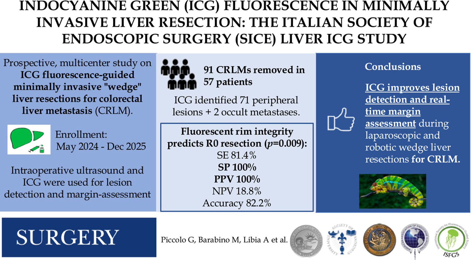
A total of 57 patients underwent minimally invasive parenchymal-sparing liver resections, removing 91 colorectal liver metastases. ICG fluorescence successfully identified 71 peripheral lesions correlating with known metastases and uncovered 2 additional subcentimeter occult metastases not detected by intraoperative ultrasound or preoperative imaging, underscoring its sensitivity.
Before resection, three fluorescence patterns were observed around lesions: rim fluorescence (the most common), partial fluorescence, and total fluorescence. These patterns likely reflect tumor-tissue interactions affecting ICG uptake and excretion.
Critically, postresection fluorescence at the margin correlated strongly with histopathologic margin status. Complete integrity of the fluorescent rim consistently predicted R0 resections in all cases. Conversely, any fluorescence irregularity—rim discontinuity, residual rim fluorescence, or protruding rim—was associated with R1 margins (positive or close margin under 1 mm). In resections where R0 was nonetheless achieved despite altered rim integrity, the margins were narrow (1 to 3 mm), indicating limited margin safety.
This real-time visual feedback allowed surgeons to identify suspicious margin areas intraoperatively potentially warranting immediate further resection to achieve clear margins.
Expert Commentary
The SICE Liver ICG study advances the field by demonstrating the practical intraoperative value of ICG fluorescence in minimally invasive liver surgery, an area where tactile feedback is limited compared to open surgery. The identification of fluorescent rim integrity as a reliable predictor for R0 resection offers a novel, objective intraoperative marker that can guide surgical decision-making.
While previous retrospective studies hinted at the utility of ICG imaging, this prospective multicenter cohort strengthens the evidence with standardized dosing, imaging protocols, and robust pathological correlation. Detecting occult lesions that evade ultrasound or preoperative imaging can potentially reduce residual disease and recurrence.
Limitations include the relatively small cohort and lack of a randomized comparator arm without fluorescence guidance. The fluorescence patterns may also vary based on tumor biology and liver function, warranting further validation across heterogeneous populations. The optimal timing and dose of ICG for maximal tumor contrast remain areas of ongoing research.
Nonetheless, this work aligns with evolving strategies prioritizing parenchymal preservation combined with precise oncological clearance. Future clinical trials should aim to standardize fluorescence assessment criteria and investigate the impact of ICG-guided resections on long-term recurrence and survival outcomes.
Conclusion
Indocyanine green fluorescence imaging is a valuable adjunct in minimally invasive, parenchymal-sparing liver resections for colorectal metastases, improving lesion detection including occult metastases and enabling real-time intraoperative margin assessment. Fluorescent rim integrity accurately predicts R0 resection status, representing a promising intraoperative biomarker for surgical precision. Integration of ICG fluorescence into liver surgery may enhance oncologic outcomes while preserving healthy liver tissue. Further standardized trials are necessary to confirm these findings and define best practices.
Funding and Registration
The study was conducted under the auspices of the Italian Society of Endoscopic Surgery (SICE). Specific funding sources were not detailed in the abstract.
References
1. Piccolo G, Barabino M, Libia A, et al. Indocyanine green (ICG) fluorescence in minimally invasive liver resection: The Italian Society of Endoscopic Surgery (SICE) liver ICG study. Surgery. 2026 Jul 16;198:110446. PMID: 42642295.
2. Ishizawa T, Fukushima N, Shibahara J, et al. Real-time identification of liver cancers by using indocyanine green fluorescent imaging. Cancer. 2009;115(11):2491-2504.
3. van der Vorst JR, Schaafsma BE, Verbeek FP, et al. Near-infrared fluorescence-guided resection of colorectal liver metastases. Cancer. 2013 Nov 1;119(21):4143-4150.
4. Peloso A, Surace S, Ceppi M, et al. Role of Indocyanine Green Fluorescence Imaging in Liver Surgery: Technological Advances and Clinical Perspectives. Cancers (Basel). 2022;14(11):2706.