Highlight
- Integration of multiplex immunofluorescence and spatial transcriptomics elucidates immune cell landscape in DLBCL.
- Identification of a high-risk subgroup with CD163high/CD11clow tumor immune microenvironment associated with poor prognosis.
- Low-risk subgroup characterized by CD163low/CD11chigh immune infiltrate correlates with favorable clinical outcomes.
- Potential therapeutic implications for checkpoint blockade with LAG3 in high-risk and CTLA4 in low-risk patients.
Study Background
Diffuse large B-cell lymphoma (DLBCL) is the most common aggressive subtype of non-Hodgkin lymphoma, characterized by considerable heterogeneity in clinical presentation and outcome. Despite advances in immunochemotherapy, including the addition of rituximab to CHOP (cyclophosphamide, doxorubicin, vincristine, and prednisone), up to 40% of patients experience relapse or refractory disease. Emerging research highlights the tumor immune microenvironment (TIME) as a critical determinant of prognosis and therapeutic response in DLBCL. Infiltrating immune cells such as macrophages and dendritic cells can influence tumor biology and modulate immune responses. However, comprehensive spatial and molecular characterization of immune cell subsets within DLBCL is limited, and their prognostic and translational relevance remains to be fully elucidated.
Study Design
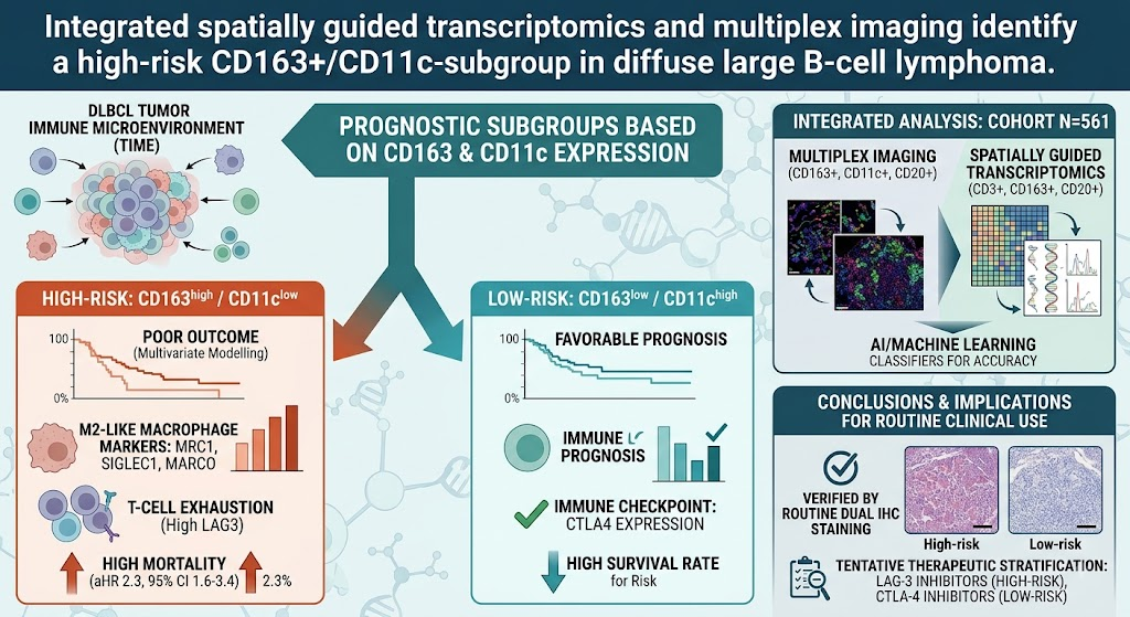
Johansson et al. conducted an integrative study combining multiplex immunofluorescence (mIF) imaging and spatially guided transcriptomics in a large cohort of 561 DLBCL patient samples. This approach enabled high-resolution spatial quantification and molecular profiling of immune and malignant cells. Key immune populations assessed included CD163-positive macrophages and CD11c-positive dendritic cells, alongside CD3-positive T cells and CD20-positive malignant B cells. Machine-learning classifiers were employed to enhance accuracy in cell density quantification. Gene expression analysis focused on immune- and cancer-related pathways to uncover underlying biological distinctions correlating with clinical outcomes. The study further validated the practicality of identifying immune subgroups using routine pathology dual immunohistochemistry (IHC) staining techniques. Clinical prognostic values were evaluated through multivariate modeling and confirmed in independent publicly available datasets comprising patients treated with CHOP and R-CHOP regimens.
Key Findings
The investigation revealed two predominant TIME subgroups with distinct prognostic implications:
- CD163low/CD11chigh Subgroup (Low Risk): This subgroup was associated with favorable prognosis. Immune profiling indicated enrichment of CD11c-positive dendritic cells and limited infiltration by CD163-positive macrophages. Notably, this TIME exhibited higher expression of the checkpoint inhibitor CTLA4, suggesting a potentially active immune surveillance environment capable of modulating antitumor responses.
- CD163high/CD11clow Subgroup (High Risk): Characterized by abundant CD163-positive macrophages, indicative of M2-like immunosuppressive macrophage phenotypes, and sparse CD11c-positive dendritic cells, this subgroup correlated with poor clinical outcomes. Transcriptomic data showed elevated expression of M2 macrophage markers such as MRC1 (mannose receptor), SIGLEC1, and MARCO. Additionally, this TIME featured T-cell exhaustion markers, particularly higher levels of LAG3, implying an immunosuppressive milieu conducive to tumor evasion.
Importantly, the prognostic significance of TIME subgroups was independent of established clinicopathological risk factors such as the International Prognostic Index (IPI). Dual IHC staining for CD163 and CD11c was demonstrated to effectively stratify patients in routine clinical settings, highlighting its translational potential. Multivariate models incorporating these markers successfully predicted patient outcomes in external cohorts treated with CHOP and R-CHOP therapies, underscoring the robustness and reproducibility of the findings.
Expert Commentary
This study elegantly leverages cutting-edge spatial transcriptomics and multiplex imaging to dissect the TIME in DLBCL at unprecedented resolution, bridging molecular phenotyping with clinically relevant prognostic stratification. The identification of a high-risk CD163high/CD11clow subgroup reinforces the detrimental role of M2-like tumor-associated macrophages, which are increasingly recognized as mediators of immune suppression and chemoresistance. Conversely, the CD11chigh signature aligns with a potentially immunostimulatory dendritic cell presence, fostering more effective antitumor immunity.
The differential expression of checkpoint molecules CTLA4 and LAG3 across subgroups offers a compelling rationale for tailored immune checkpoint inhibitor therapies. LAG3 blockade may revive exhausted T cells in the high-risk group, whereas targeting CTLA4 could enhance antitumor immunity in the low-risk group. However, prospective clinical trials are necessary to validate these hypotheses and assess therapeutic efficacy in DLBCL subpopulations stratified by TIME profiling.
Limitations include potential heterogeneity within analyzed immune subsets and the cross-sectional nature of tissue sampling. Furthermore, functional studies are needed to precisely define mechanistic pathways through which these immune cell phenotypes influence lymphoma behavior and treatment response.
Conclusion
This comprehensive spatial and transcriptomic analysis defines clinically relevant immune microenvironment subgroups in DLBCL with independent prognostic value beyond conventional risk factors. The demonstration of a high-risk CD163+/CD11c- phenotype associated with immune exhaustion and poor outcome paves the way for biomarker-driven therapeutic stratification. Integration of CD163 and CD11c dual IHC staining into routine pathological assessment offers a feasible method to guide prognosis and potentially personalize checkpoint immunotherapy strategies in DLBCL. Future research should focus on validating these findings prospectively and exploring targeted immunomodulatory treatments tailored to these TIME-defined patient subsets.
Funding and Clinical Trial Registration
Details on funding sources and clinical trial registrations were not specified in the original publication.
References
- Johansson A, Nilsson D, Wikström F, et al. Integrated spatially guided transcriptomics and multiplex imaging identify a high-risk CD163+/CD11c-subgroup in diffuse large B-cell lymphoma. Haematologica. 2026 Jul 23. PMID: 42489074.
- Scott DW, Gascoyne RD. The tumour microenvironment in B cell lymphomas. Nat Rev Cancer. 2014 Sep;14(9):517-34. doi: 10.1038/nrc3775.
- Havel JJ, Chowell D, Chan TA. The evolving landscape of biomarkers for checkpoint inhibitor immunotherapy. Nat Rev Cancer. 2019 Sep;19(3):133-150. doi: 10.1038/s41568-019-0116-x.
- Binnewies M, Roberts EW, Kersten K, et al. Understanding the tumor immune microenvironment (TIME) for effective therapy. Nat Med. 2018 May;24(5):541-550. doi: 10.1038/s41591-018-0014-x.