Highlight
– Modified focal excision technique based on pathoanatomical defects and neovascularization offers uterus preservation in placenta percreta.
– The technique shows high feasibility with a 93% success rate in uterus preservation in 15 cases.
– Median blood loss was 2000 mL with manageable morbidity, including transient bladder injuries.
– Approach allows targeted placental resection, avoiding cesarean hysterectomy in most cases.
Study Background
Placenta Accreta Spectrum (PAS) disorders represent a spectrum of abnormal placental adherence and invasion into the uterine wall, ranging from placenta accreta to the more severe placenta percreta, in which placental tissue penetrates beyond the uterine serosa, often involving adjacent organs like the bladder. PAS incidence has risen with increasing cesarean sections and advanced maternal age, posing significant maternal morbidity and mortality risks due to massive hemorrhage and complex surgical management.
Cesarean hysterectomy remains the standard approach for severe PAS cases but carries high morbidity, including hemorrhage, bladder injury, and irreversible loss of fertility. Conservative and uterus-preserving techniques are of increasing interest, aiming to maintain fertility and reduce surgical risks. However, these are challenging, especially in placenta percreta, where neovascularization and extensive abnormal placental infiltration complicate resection.
This study by Vural et al. assesses a modified focal excision strategy for PAS, focusing on pathoanatomical defects and the pattern of neovascularization rather than solely on the depth of tissue invasiveness. The objective is to determine the feasibility, safety, and outcomes of this uterus-sparing approach using a standardized surgical protocol.
Study Design
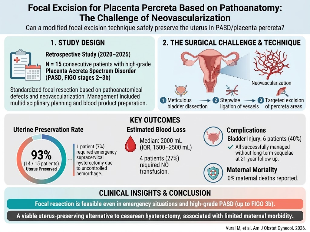
A retrospective review was conducted on 15 consecutive patients diagnosed with PAS stages 2 to 3b according to International Federation of Gynecology and Obstetrics (FIGO) criteria, treated between 2020 and 2025 at a tertiary referral center. Diagnosis involved abdominal and transvaginal ultrasonography confirming PAS severity, supplemented by multidisciplinary assessment and detailed patient counseling on risks and benefits.
Preoperative planning included preparation for potential massive transfusion and coordination among obstetrics, anesthesiology, urology, and critical care teams. The surgical approach utilized a standardized focal resection technique targeting abnormal placental areas identified intraoperatively through defect mapping and vascular patterns.
The procedure includes meticulous bladder dissection, stepwise ligation of feeding vessels, and excision of placental tissue limited to the percreta focal points, sparing unaffected uterine tissue. Intraoperative video documentation and postoperative histopathology verified the targeted excision and confirmed diagnosis.
Key Findings
Uterus preservation was achieved in 14 out of 15 patients (93%), demonstrating high technique feasibility even in advanced PAS grades (up to FIGO 3b). One patient required emergency supracervical hysterectomy due to uncontrolled hemorrhage, reflecting the procedure’s limitation in rare severe bleeding episodes.
The median estimated blood loss was 2000 mL (interquartile range 1500–2500 mL), with four patients not requiring any blood transfusion, highlighting effective hemorrhage control. Although surgical challenges included bladder injury in 6 patients (40%), all injuries were successfully managed intraoperatively or postoperatively without long-term complications during follow-up exceeding one year.
No maternal deaths occurred in this series, an encouraging safety indicator when compared with traditional cesarean hysterectomy data, which often report higher morbidity and mortality risks.
The focal excision method was also feasible in emergency settings, allowing rapid yet precise surgical management of hemorrhage. The correlation between intraoperative findings, video recordings, and histopathology confirmed reproducibility and diagnostic accuracy.
Expert Commentary
Current guidelines generally recommend cesarean hysterectomy for placenta percreta to control life-threatening hemorrhage; however, this approach leads to loss of fertility and high complication rates. This study challenges the traditional paradigm by leveraging anatomical and vascular insights to inform a conservative resection technique.
Neovascularization in PAS contributes to surgical complexity by creating fragile and aberrant blood vessel networks that increase bleeding risk. By dissecting and ligating feeding vessels stepwise and focusing excision on placental defects, the authors propose a rational method to mitigate hemorrhage without radical uterine removal.
Nonetheless, the procedure demands advanced surgical expertise, multidisciplinary collaboration, and thorough preoperative planning. The notable bladder injury rate indicates inherent procedural risk, warranting experienced surgeons and close follow-up.
Limitations include the retrospective design, small sample size, and single-center experience. Larger prospective cohorts and randomized studies are necessary to validate safety and long-term fertility outcomes.
Conclusion
This clinical series supports the feasibility and safety of a pathoanatomy-based focal excision technique for managing placenta percreta, achieving high uterus preservation rates and acceptable morbidity. The approach offers a promising uterus-sparing alternative to cesarean hysterectomy in carefully selected patients, addressing an unmet need for fertility-preserving PAS treatments.
Future research should explore standardized protocols, long-term reproductive outcomes, and comparative effectiveness against traditional surgical methods to guide practice integration.
Funding and ClinicalTrials.gov
The published article does not report specific funding sources or clinical trial registration numbers.
References
1. Vural M, Pohle-Rüffer MC, Rauh M, Kappelmeyer M, Köninger A. Focal excision for placenta percreta based on pathoanatomy: the challenge of neovascularization. Am J Obstet Gynecol. 2026 May 19;235(4):943-951. PMID: 42162710.
2. Committee on Obstetric Practice. Committee Opinion No. 778: Placenta Accreta Spectrum. Obstet Gynecol. 2019;133(1):e91-e98.
3. Jauniaux E, Bhide A, Kennedy A, Woodward P, Hubinont C, Collins S. FIGO classification for the clinical diagnosis of placenta accreta spectrum disorders. Int J Gynaecol Obstet. 2019 Dec;146(2):20-24.
4. Sentilhes L, Kayem G, Ambroselli C, et al. Conservative management of placenta percreta: a multicenter study. Obstet Gynecol. 2010;115(3):505-514.