Highlight
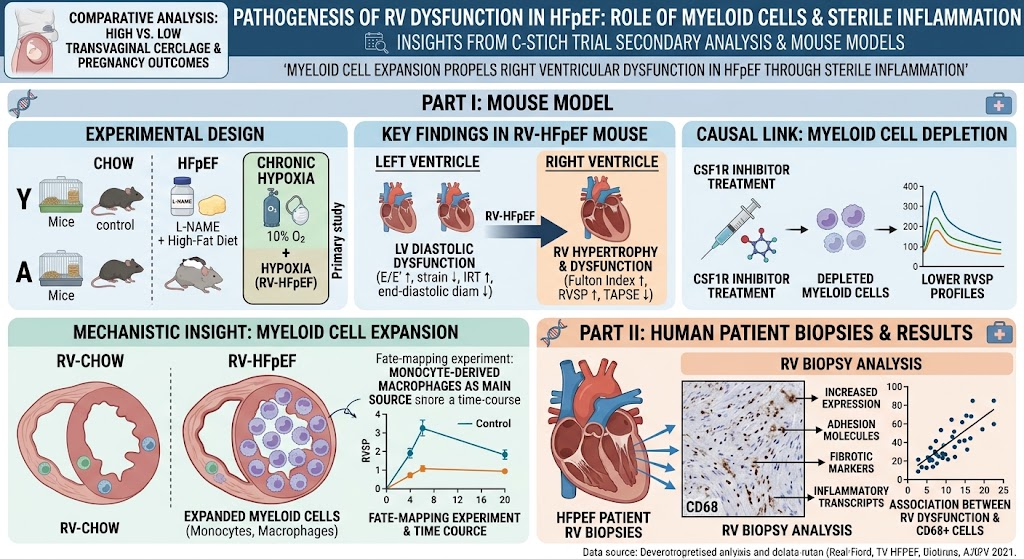
- Right ventricular dysfunction (RVD) in heart failure with preserved ejection fraction (HFpEF) is linked to immune cell infiltration, primarily involving myeloid lineage cells.
- A novel murine model combining HFpEF induction with chronic hypoxia replicates clinically relevant RV dysfunction and myeloid cell expansion.
- Myeloid cell depletion ameliorates RV pressure overload, underscoring the causal role of sterile inflammation in RV remodeling.
- Human HFpEF RV biopsies show increased inflammatory and fibrotic markers, correlating immune cell presence with RVD severity.
Study Background
Heart failure with preserved ejection fraction (HFpEF) is a prevalent and multifactorial cardiovascular syndrome characterized by symptoms of heart failure despite normal left ventricular systolic function. Increasing evidence highlights the importance of right ventricular dysfunction (RVD) as a key determinant of morbidity and mortality in HFpEF patients. However, the mechanisms driving RVD in this subgroup remain insufficiently understood. Emerging data suggest that inflammation and immune cell-mediated processes may play a role, but lack of suitable small animal models with manifest RVD has limited mechanistic exploration. Understanding how leukocyte dynamics influence RV remodeling could reveal novel therapeutic targets, addressing a major unmet need in HFpEF management.
Study Design
This investigation employed a translational research approach combining advanced murine modeling with human tissue analysis. Male and female C57BL/6J mice, stratified by age into young (80 weeks) cohorts, were assigned to four experimental conditions: control chow diet, HFpEF induction via L-NAME combined with a high-fat diet, chronic hypoxia exposure (10% O2), and a combined HFpEF plus hypoxia group (RV-HFpEF). Comprehensive assessment of biventricular function was performed using echocardiography and hemodynamics. Myeloid cell dynamics were evaluated through flow cytometry and proteomic profiling of ventricular tissues. To establish causality, the RV-HFpEF group underwent intervention with a colony-stimulating factor 1 receptor (CSF1R) inhibitor to deplete myeloid cells. Additionally, right ventricular biopsies from human HFpEF patients were analyzed for inflammatory markers, fibrosis, and immune cell infiltration.
Key Findings
The RV-HFpEF model reproduced hallmark features of HFpEF-associated right ventricular dysfunction. Compared to controls, RV-HFpEF mice displayed left ventricular diastolic dysfunction indicated by elevated E/E’ ratios and impaired global longitudinal peak strain, along with smaller end-diastolic diameters and prolonged isovolumetric relaxation time. RV remodeling manifested as significant hypertrophy with increased Fulton index and elevated RV systolic pressure, accompanied by impaired RV systolic function evidenced by reduced tricuspid annular plane systolic excursion.
Leukocyte profiling revealed pronounced expansion of total leukocytes, monocytes, and macrophages selectively in RV tissue of RV-HFpEF mice. Proteomic analysis confirmed enrichment of proteins involved in innate immune activation, macrophage chemotaxis, and leukocyte migration within the RV. Notably, fate-mapping indicated that recruited monocyte-derived macrophages became the predominant macrophage population in the RV during disease progression. Intervention with a CSF1R inhibitor effectively depleted myeloid cells and resulted in lower RV systolic pressure compared to untreated RV-HFpEF mice, demonstrating a direct pathogenic contribution of these immune cells to RV pressure overload and dysfunction.
Translational relevance was demonstrated through analysis of human RV biopsies from HFpEF patients. These samples exhibited elevated expression of adhesion molecules, pro-fibrotic markers, and inflammatory transcripts. Furthermore, abundance of CD68+ macrophages correlated with severity of RVD, supporting a role for sterile inflammation mediated by myeloid cells in human HFpEF pathophysiology.
Expert Commentary
This rigorous study fills a critical gap in HFpEF research by elucidating the immunopathogenic mechanisms underpinning RV dysfunction—a core driver of adverse outcomes. The innovative use of a combined HFpEF plus hypoxia murine model faithfully replicates the clinical scenario of RV pressure overload and dysfunction, enabling mechanistic dissection. The identification of monocyte-derived macrophages as key effectors indicates that targeted modulation of myeloid cell recruitment or activation may represent a novel therapeutic avenue.
While the animal model provides invaluable insight, translation to human disease will require further validation in diverse HFpEF phenotypes and exploration of long-term effects of immunomodulation. Moreover, sterile inflammation likely interacts with other pathophysiological processes such as metabolic derangements and neurohormonal activation, meriting integrative investigation.
Conclusion
Dysregulated myeloid cell dynamics drive right ventricular dysfunction in HFpEF through mechanisms of sterile inflammation. This work underscores the pivotal role of the innate immune system in cardiac remodeling beyond the left ventricle and highlights myeloid cells as promising targets for intervention. Future research should aim to validate these findings in clinical trials and explore therapeutic strategies to ameliorate RV dysfunction and improve outcomes in HFpEF.
Funding and Disclosure
The study was supported by multiple institutional and research grants as listed in the original publication. No conflicts of interest were reported.
References
1. Jaeschke L, Koçana C, Chitroceanu AM, et al. Myeloid Cell Expansion Propels Right Ventricular Dysfunction in HFpEF Through Sterile Inflammation. Circulation. 2026; PMID: 42610249.
2. Shah SJ. The pathophysiology of HFpEF: insights and questions. Heart Fail Clin. 2023;19(3):xx-xx.
3. van Heerebeek L, Paulus WJ. Myocardial microvascular inflammation in HFpEF. Heart Fail Rev. 2019;24(2):195–203.