Highlight
1. Higher fat-free mass (FFM) levels alone may misleadingly suggest increased metabolic risk unless accounted for ectopic fat compartments.
2. Visceral adipose tissue (VAT) prominently modifies the relationship between FFM and metabolic syndrome prevalence.
3. Muscle fat infiltration (MFI) also influences the FFM-metabolic outcomes linkage, partially independent of VAT.
4. Circulating sex hormones show modest modifying effects, varying by the specific FFM metric employed.
Study Background
Fat-free mass (FFM), encompassing muscle mass and other non-fat components, has traditionally been viewed as synonymous with skeletal muscle—an essential organ for glucose uptake and insulin sensitivity. Skeletal muscle mass is often inversely correlated with metabolic syndrome (MetS), a cluster of conditions increasing cardiovascular and diabetic risk. However, emerging evidence from contemporary large cohorts has challenged the simplistic interpretation of FFM by revealing paradoxical associations of higher FFM with insulin resistance and adverse metabolic profiles. This complexity may arise because FFM metrics often fail to distinguish between metabolically active muscle and infiltrative or ectopic fat within muscle, as well as visceral adipose deposits. Accurately interpreting the role of FFM in metabolic health thus necessitates a detailed understanding of the heterogeneity within fat-free compartments and the influence of hormonal milieu.
Study Design
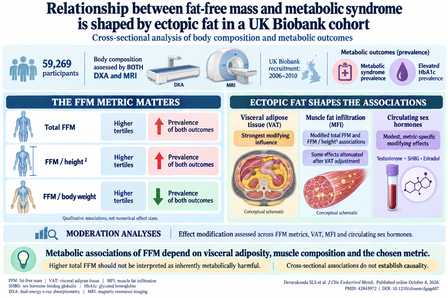
This cross-sectional investigation utilized data from the UK Biobank, enrolling 59,269 participants collected between 2006 and 2010. Both dual-energy X-ray absorptiometry (DXA), to quantify total and regional body composition, and magnetic resonance imaging (MRI), to assess ectopic fat depots including visceral adipose tissue (VAT) and muscle fat infiltration (MFI), were employed — offering comprehensive phenotyping of fat distribution and quality of fat-free tissues. The study examined three metrics of fat-free mass: total FFM, FFM adjusted to height squared (FFM/height2), and proportion of FFM relative to body weight (FFM/Wt). Outcomes of interest included prevalence of metabolic syndrome (MetS) and elevated glycated hemoglobin (HbA1c), a marker of glucose dysregulation. Moderation analyses assessed the modifying effects of VAT, MFI, and circulating sex hormones—testosterone, sex hormone-binding globulin (SHBG), and estradiol—on the associations between FFM and metabolic parameters.
Key Findings
The study revealed that the relationship between FFM and metabolic outcomes varies considerably depending on how FFM is expressed and the degree of ectopic fat present:
- FFM expressed as total mass and adjusted for height (FFM/height2) showed a direct association with adverse metabolic outcomes. Specifically, higher tertiles of total FFM and FFM/height2 were associated with increased prevalence of metabolic syndrome and elevated HbA1c levels, contrary to the traditional conception that more muscle mass alone is metabolically protective.
- Conversely, higher FFM relative to body weight (FFM/Wt) exhibited an inverse association with these metabolic risk markers. This suggests that when accounting for overall body size and fatness, a greater relative fat-free component corresponds to better metabolic health.
- Visceral adipose tissue emerged as the strongest modifier in these relationships. Increased VAT substantially amplified the adverse metabolic risks associated with higher absolute FFM metrics. This supports the concept that concomitant ectopic fat in visceral compartments negates the beneficial metabolic impact of increased muscle or fat-free mass.
- Muscle fat infiltration also modulated FFM-metabolic syndrome associations. Higher MFI correlated with higher metabolic risk independent of overall muscle mass, although its effect was partially attenuated after adjusting for VAT, indicating interplay between various fat depots within and around muscle tissue.
- Sex hormones exerted modest yet statistically significant modifying effects. Testosterone, SHBG, and estradiol influenced the associations in metric-specific manners, reflecting their biological roles in muscle maintenance, fat distribution, and insulin sensitivity. However, their impact was less pronounced than that of ectopic fat compartments.
Expert Commentary
This robust, well-powered cross-sectional study challenges the conventional straightforward interpretation of fat-free mass as an unequivocal marker of metabolic health. The findings underscore the critical importance of considering ectopic fat deposits—particularly VAT and intra-muscular fat—when assessing the metabolic implications of muscle mass or FFM. The differential associations observed depending on the FFM metric chosen provide methodological insight; FFM relative to weight may be a more physiologically meaningful measure internally adjusting for fatness compared to raw or height-adjusted FFM.
These observations align with prior mechanistic research demonstrating that ectopic fat exerts pro-inflammatory and insulin resistance–promoting effects within muscle and viscera, complicating the metabolic landscape of individuals ostensibly possessing robust lean mass. Furthermore, subtle hormonal influence highlights the need for nuanced endocrine assessment when evaluating body composition and metabolic risk, particularly in men and women with varying sex hormone profiles.
Nevertheless, as a cross-sectional analysis, causality cannot be inferred, and residual confounding is inevitable despite rigorous statistical adjustments. The generalizability to ethnically diverse or clinical populations requires further validation, and longitudinal studies are warranted to investigate temporal changes in fat depots, FFM, and metabolic outcomes.
Conclusion
This study elucidates that the metabolic risk traditionally attributed to fat-free mass is not intrinsic to FFM itself but is substantially shaped by ectopic adiposity—primarily visceral fat and muscle fat infiltration—and the method of FFM quantification. Clinicians and researchers should exercise caution in interpreting FFM values without accounting for these modifying factors. Understanding this complex interplay may improve metabolic risk stratification, guide personalized interventions targeting fat redistribution, and refine body composition assessments in metabolic syndrome management and research.
Funding and ClinicalTrials.gov
The original study was conducted using data from the UK Biobank resource. No specific funding information or clinical trials registration was provided in the source citation.
References
1. Devarakonda SLS, Zhang D, Roberts R, Rebello C. Relationship between fat-free mass and metabolic syndrome is shaped by ectopic fat in a UK Biobank cohort. J Clin Endocrinol Metab. 2026 Oct 8; PMID: 42843972.
2. Srikanthan P, Karlamangla AS. Muscle mass index as a predictor of mortality in older adults. J Gerontol A Biol Sci Med Sci. 2014; 69(9):1095-1100.
3. Tchoukalova YD, Votruba SB, Tchkonia T, et al. Regional adipose tissue distribution and insulin resistance. Yale J Biol Med. 2006; 79(3-4):125-130.
4. Goodpaster BH, Krishnaswami S, Harris TB, et al. Obesity, regional body fat distribution, and the metabolic syndrome in older men and women. Arch Intern Med. 2005;165(7):777-783.
5. Kelley DE, Goodpaster BH, Wing RR, Simoneau JA. Skeletal muscle triglyceride level is inversely related to insulin action. Diabetes. 1999;48(6): 1329-1334.
6. Wildman RP, Herber-Gast G, Koczwara K, et al. Associations of visceral fat and ectopic fat with insulin resistance and cardiometabolic risk. J Clin Endocrinol Metab. 2022;107(4): e1421–e1430.
7. Bhasin S, Woodhouse L, Storer TW. Testosterone and anabolic steroids in endocrine function. J Clin Endocrinol Metab. 2001;86(11):541-555.