Highlight
- Mid-phase hyperfluorescent plaques (MPHPs) on ICGA exhibit distinct phenotypic stages correlating with structural retinal changes on OCT in CSCR.
- Ellipsoid zone/retinal pigment epithelium (EZ/RPE) irregularity is nearly universal across all MPHP stages and may represent an underlying susceptible feature in CSCR.
- Progression of MPHPs correlates strongly with increased subretinal fluid (SRF), hyperreflective foci (HRF), pigment epithelial detachment (PED), and outer nuclear layer (ONL) thinning on OCT.
- MPHPs act as dynamic focal sites of disease activity within the hyperpermeable choroid, informing potential targets for monitoring and therapeutic intervention.
Study Background
Central serous chorioretinopathy (CSCR) is a common retinal disorder characterized by serous detachment of the neurosensory retina due to leakage at the level of the retinal pigment epithelium (RPE). The pathophysiology is closely tied to choroidal hyperpermeability and RPE dysfunction, but precise lesion characterization remains challenging. Indocyanine green angiography (ICGA) is instrumental for visualizing choroidal vasculature and identifying hyperfluorescent plaques that signify focal changes in choroidal permeability. However, the phenotypic spectrum of these mid-phase hyperfluorescent plaques (MPHPs) and their corresponding optical coherence tomography (OCT) features are incompletely understood, limiting comprehensive disease assessment and management. This study addresses this knowledge gap by applying multimodal imaging to characterize MPHPs in CSCR and explore their clinical significance.
Study Design
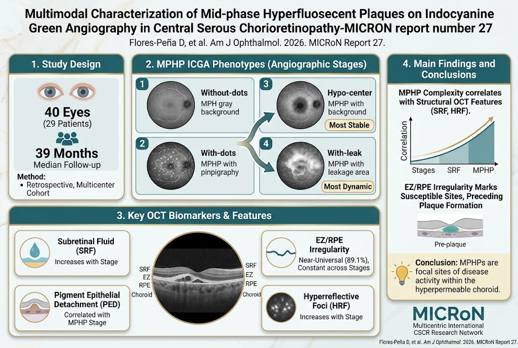
This retrospective, observational multicenter cohort study included 40 eyes from 29 CSCR patients with a median follow-up of 39 months. The patient cohort was predominantly male (86.2%), reflecting known CSCR epidemiology. Baseline and follow-up imaging data were systematically analyzed using ICGA to identify and classify MPHPs into four distinct types: ‘without-dots’, ‘with-dots’, ‘hypo-center’, and ‘with-leak’. When plaques exhibited multiple coexisting features, a hierarchical ‘stage’ was assigned according to the highest-ranking angiographic attribute. Corresponding OCT features at MPHP sites were meticulously assessed for structural correlates including subretinal fluid (SRF), ellipsoid zone and RPE irregularities (EZ/RPE), pigment epithelial detachment (PED), hyperreflective foci (HRF), outer nuclear layer (ONL) thinning, double-layer sign, RPE atrophy, RPE infrared abnormalities, pachyvessels, and choroidal thickness. Statistical associations were determined using generalized estimating equations with adjustments for multiple comparisons. Transitions between plaque stages and predictors for new leakage onset were also analyzed longitudinally.
Key Findings
At baseline, 112 MPHPs were documented, with 92 plaques having co-registered OCT scans. EZ/RPE irregularity was near-universal, observed in approximately 89.1% of plaques, and was evenly distributed across all plaque stages without significant variation. This suggests EZ/RPE irregularity is an early, stable marker of plaque susceptibility rather than representing active progression.
The presence of subretinal fluid (SRF) demonstrated the strongest association with advanced plaque stages, culminating in 100% prevalence in the ‘with-leak’ stage. Odds ratio (OR) for SRF in higher-ranking stages was 5.34 (95% CI: 2.90-9.84; q <0.001), indicating a robust correlation between angiographic activity and fluid accumulation.
Additional OCT features such as hyperreflective foci (HRF) showed an OR of 1.66 (q <0.001), pigment epithelial detachment (PED) an OR of 1.99 (q=0.019), and outer nuclear layer thinning an OR of 1.93 (q=0.039), all progressively more common in advanced plaque stages. These structural changes reflect escalating retinal and RPE disruption concurrent with angiographic complexity.
Among sites where new MPHPs emerged during follow-up, EZ/RPE irregularity was already present in 8 of 10 assessable locations prior to plaque appearance, whereas other OCT abnormalities typically were not. This finding positions EZ/RPE irregularity as a possible precursor lesion indicative of focal vulnerability.
Dynamic plaque stage transitions were observed, with the most common shift from ‘with-dots’ to ‘hypo-center’. Plaques categorized as ‘hypo-center’ were notably stable, remaining unchanged in 78.3% of cases and exhibiting the lowest progression rates to more advanced stages.
Expert Commentary
This study robustly delineates the structural and angiographic evolution of MPHPs in CSCR, underscoring their complex, dynamic nature. The persistent presence of EZ/RPE irregularities regardless of plaque stage suggests these changes may constitute a primary site of choroidal-RPE susceptibility, potentially critical in CSCR pathogenesis. The strong correlation between plaque stage and accumulation of subretinal fluid and other structural OCT abnormalities provides insight into disease activity and progression.
These findings can assist clinicians in prognostication and tailoring monitoring strategies, emphasizing the utility of multimodal imaging to detect early vulnerable sites before overt leakage ensues. Limitations include the retrospective design and restriction to imaging biomarkers without parallel functional correlation; prospective validation assessing visual outcomes will further clarify clinical implications.
Conclusion
Mid-phase hyperfluorescent plaques on ICGA represent focal, dynamic sites of disease activity in CSCR, with escalating structural disruption evident on OCT correlating with increasing plaque complexity. EZ/RPE irregularity emerges as a key early and stable marker of plaque presence and potential leakage risk. Multimodal imaging-based phenotyping of MPHPs adds valuable granularity to CSCR evaluation, fostering improved understanding, monitoring, and potentially targeted intervention in this enigmatic chorioretinopathy.
Funding and ClinicalTrials.gov
The study was conducted by the Multicentric International CSCR Research Network (MICRoN). Funding details were not explicitly provided in the original report. There is no mention of a ClinicalTrials.gov registration number.
References
1. Flores-Peña D, et al. Multimodal Characterization of Mid-phase Hyperfluorescent Plaques on Indocyanine Green Angiography in Central Serous Chorioretinopathy- MICRoN report number 27. Am J Ophthalmol. 2026 Oct 1; PMID: 42822586.
2. Spaide RF, et al. Central Serous Chorioretinopathy. Retina. 2018;38(10):1986-1993.
3. Daruich A, Matet A, Dirani A, et al. Central Serous Chorioretinopathy: Recent Findings and New Physiopathology Hypothesis. Prog Retin Eye Res. 2015 Sep;48:82-118.
4. Bousquet E, et al. Optical coherence tomography angiography of type I choroidal neovascularization in chronic central serous chorioretinopathy. Am J Ophthalmol. 2017;178:68–77.