
Highlight
This study validates preoperative ultrasound as a precise, reproducible modality to localize lymphovenous anastomosis (LVA) sites, showing a positive predictive value (PPV) of 95.4% against intraoperative confirmation. Ultrasound measurements closely corresponded with intraoperative vessel sizes, and procedural efficiency was practical, with an average examination time under 35 minutes. This technique demonstrated consistent effectiveness regardless of patient or anatomical variables, positioning ultrasound as a frontline tool in supermicrosurgical planning for lymphedema treatment.
Study Background
Lymphedema is a chronic, often debilitating condition characterized by lymphatic fluid accumulation leading to limb swelling, fibrosis, and functional impairment. Lymphovenous anastomosis (LVA) is a supermicrosurgical intervention aiming to restore lymphatic drainage by creating connections between lymphatic vessels and adjacent veins. Successful LVA surgery critically depends on preoperative identification and mapping of suitable lymphatic and venous structures.
Traditional lymphatic imaging relies heavily on indocyanine green (ICG) lymphography, which while widely used, suffers limitations such as poor depth penetration and reduced visibility in advanced disease stages. There is an unmet need for alternative or complementary imaging modalities capable of improving surgical planning accuracy and outcomes in lymphedema care.
Study Design
This investigation is a retrospective analysis of prospectively collected data from April 2019 to January 2024 in a secondary care setting. The study cohort included 87 patients (mean age 55.8 ± 12.8 years, predominantly female) with scintigraphically confirmed limb lymphedema. All patients underwent preoperative ultrasound examinations aimed at identifying potential LVA sites. The intraoperative identification of lymphatic and venous anatomy served as the gold standard reference for evaluating the diagnostic accuracy of ultrasound localization.
Diagnostic performance measures included the positive predictive value (PPV) of ultrasound-identified sites selected for surgery, Bland-Altman assessments comparing ultrasound-determined vessel diameters with intraoperative measurements, and logistic regression analyses exploring predictors of false positives. Efficiency metrics such as total examination time and per-image analysis time were also recorded.
Key Findings
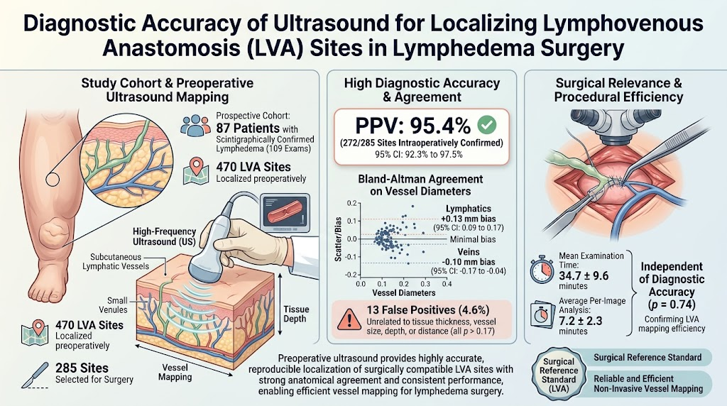
From 109 ultrasound examinations encompassing 470 potential LVA sites, 285 sites were chosen for intraoperative exploration. Of these, 272 sites were confirmed as surgically compatible vessels, yielding a high PPV of 95.4% (95% CI: 92.3, 97.5%). Only 13 sites identified by ultrasound were falsely positive, and these false positives did not statistically correlate with tissue thickness, vessel size, depth, or vessel distance, indicating robust performance across diverse anatomical variables.
Diameter measurements derived from ultrasound demonstrated minimal bias relative to intraoperative findings. Bland-Altman analysis revealed a slight overestimation bias of +0.13 mm for lymphatic vessels and a slight underestimation bias of -0.10 mm for veins, both well within clinically acceptable limits.
The average total ultrasound examination lasted 34.7 ± 9.6 minutes, with individual image analysis averaging 7.2 ± 2.3 minutes. Importantly, the time spent analyzing images was not predictive of diagnostic accuracy, suggesting procedural efficiency does not compromise the reliability of vessel localization.
The study highlights that ultrasound can effectively overcome the depth penetration limitations of ICG lymphography, especially benefiting patients with advanced lymphedema where visualization is challenging.
Expert Commentary
Preoperative mapping of lymphatic and venous vessels is indispensable for the success of LVA surgeries, which demand identification of vessels as small as 0.5 mm in diameter. While ICG lymphography remains the standard, its limitations in deeper or fibrotic tissues create challenges in advanced cases. This study provides compelling evidence supporting ultrasound as a complementary or alternative imaging approach.
Ultrasound’s ability to render cross-sectional anatomical details in real-time facilitates precise measurement and localization of lymphatic and venous structures. The high positive predictive value and strong measurement agreement with intraoperative data underscore its utility. Additionally, the independence of diagnostic accuracy from tissue characteristics could simplify patient selection and surgical planning.
However, ultrasound is operator-dependent and requires specific expertise, especially in supermicrosurgical contexts. Broader implementation would necessitate training programs and standardization efforts to ensure reproducibility across centers.
Limitations of the study include its retrospective nature and potential selection bias inherent in single-center analyses. Further multicenter, prospective trials comparing ultrasound directly with established modalities like ICG lymphography and lymphoscintigraphy would help clarify the optimal imaging algorithm.
Conclusion
Ultrasound presents a robust, accurate, and efficient method for preoperative localization of lymphovenous anastomosis sites in lymphedema surgery. Its high diagnostic accuracy and measurement concordance with intraoperative findings support its integration into surgical planning protocols. By addressing limitations of current imaging techniques, ultrasound can potentially improve surgical outcomes and expand treatment options for patients with advanced lymphedema.
Future directions should focus on technology refinement, standardized imaging protocols, and multicenter validation to optimize utilization in clinical practice.
Funding and Clinical Trials
The article as referenced does not specify funding sources or clinical trial registration numbers. Further details would be available from the original publication.
References
1. Jaklin FJ, Oberreiter ML, Weninger WJ, Tzou CJ, Meng S. Diagnostic Accuracy of Ultrasound for Localizing Lymphovenous Anastomosis Sites in Lymphedema Surgery. Plastic and Reconstructive Surgery. 2026 Oct 14; PMID: 42878748.
2. Damstra RJ, Leavesley DI, Taylor GI. Lymphaticovenous anastomosis for treatment of lymphedema: a review of techniques. J Reconstr Microsurg. 2016;32(5):360-372.
3. Unno N, Inuzuka K, Suzuki M, Yamamoto N, Sagara D, Nishiyama M, Konno H. The earliest change in lymphatic function detected by indocyanine green lymphography in asymptomatic limbs of lower extremity lymphedema patients. J Vasc Surg Venous Lymphat Disord. 2015;3(4):403-410.
4. Szuba A, Shin WS, Strauss HW, Rockson SG. The third circulation: radionuclide lymphoscintigraphy in the evaluation of lymphedema. J Nucl Med. 2003;44(1):43-57.