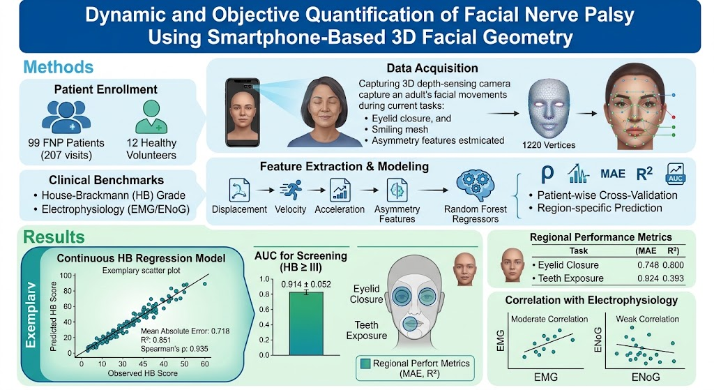
Highlight
This study introduces a scalable, smartphone-based method to objectively quantify facial nerve palsy (FNP) severity using high-resolution 3D facial geometry and advanced machine learning algorithms, achieving accurate side-aware House-Brackmann (HB) grading and state-of-the-art clinical screening.
The continuous HB score prediction using displacement, velocity, acceleration, and asymmetry features attained high correlations (Spearman’s ρ=0.935) and low error rates, surpassing human grading resolution and detecting subtle residual asymmetry.
Region-specific analyses showed optimal performance in assessing eyelid closure and teeth exposure, critical functional parameters in FNP evaluation.
This approach has strong potential to facilitate early diagnosis, standardized longitudinal monitoring, and home-based rehabilitation of FNP patients.
Study Background
Facial nerve palsy (FNP), characterized by unilateral facial muscle weakness, poses significant morbidity, impacting patients’ facial expression, communication, and psychosocial well-being. Clinical evaluation relies primarily on subjective grading scales like the House-Brackmann (HB) scale, which are limited by inter-rater variability and categorical rather than continuous assessment.
Electrophysiologic tests such as electroneurography and electromyography provide objective measures, but are invasive, technically demanding, and not easily repeated for frequent monitoring. There is an unmet need for accessible, quantitative, and reproducible tools that allow dynamic, continuous, and side-specific assessment of FNP severity to optimize personalized care and rehabilitation outcomes.
Recent advances in smartphone 3D front-facing cameras allow high-frame-rate capture of detailed facial geometry in three dimensions, which could revolutionize FNP evaluation by providing objective, low-cost, and patient-centered monitoring outside clinical settings.
Study Design
This prospective study enrolled 99 patients with peripheral acute unilateral FNP over 207 clinical visits, alongside 12 healthy volunteers, between July 2021 and February 2024. Facial dynamics were recorded using a smartphone equipped with a 3D front-facing camera, generating a dense facial mesh of 1220 three-dimensional vertices.
Multiple standardized facial tasks were recorded, including eyelid closure and teeth exposure, to capture different regions of facial movement. Clinical benchmarks included the HB grading scale and electrophysiological measures such as integrated electromyography and electroneurography.
Extracted features included quantitative parameters of facial vertex displacement, velocity, acceleration, and asymmetry between affected and unaffected sides. Machine learning models—random forest regressors—were trained to predict a signed continuous HB score that accounted for the side of palsy, as well as region-specific scores.
Patient-wise cross-validation ensured robustness of predictions. Performance metrics used were Spearman’s correlation coefficient (ρ), mean absolute error (MAE), coefficient of determination (R2), and area under the receiver operating characteristic curve (AUC) for detection of clinically relevant palsy (HB ≥ III).
Key Findings
The continuous HB regression model demonstrated excellent predictive performance with a MAE of 0.718, R2 of 0.851, and a very strong correlation (Spearman’s ρ=0.935), indicating high accuracy and precision of the model in estimating facial palsy severity on a continuous scale.
Predictions in healthy controls clustered tightly around normal values, confirming the method’s robustness to false-positive detection. For clinical screening aimed at detecting significant palsy (HB ≥ III), the model achieved an AUC of 0.914 ± 0.052, indicating excellent diagnostic discrimination.
Region-specific analyses highlighted that assessments focusing on eyelid closure and teeth exposure yielded the best predictive accuracy, underscoring the functional importance of these regions in FNP severity assessment and their suitability for objective monitoring.
Comparisons to electrophysiological tests showed moderate correlations with integrated electromyography but weaker correlations with electroneurography, suggesting that 3D facial geometry captures functional impairments complementary to electrophysiological signals.
Expert Commentary
This study addresses a critical gap in FNP management by providing an accessible, objective, and dynamic quantification tool leveraging readily available smartphone technology. The continuous, side-aware HB score prediction reduces the ceiling effects and subjectivity inherent in traditional ordinal scales, facilitating more nuanced monitoring of disease progression and recovery.
Integration of motion dynamics such as velocity and acceleration alongside asymmetry enhances the model’s sensitivity to subtle facial dysfunctions often imperceptible to clinicians, potentially improving early detection and customized rehabilitation.
Limitations include the need to validate this approach across diverse populations, varying smartphone hardware, and in chronic or bilateral FNP cases. Additionally, the dependency on patient compliance and technical factors such as lighting and positioning requires consideration for clinical deployment.
Conclusion
Smartphone-based high-frame-rate 3D facial geometry capture, combined with advanced machine learning, provides an innovative and clinically practical method for objective, dynamic, and continuous quantification of facial nerve palsy severity.
This technology offers high accuracy in side-specific HB grading and excellent performance in screening for clinically meaningful palsy, with potential applications in early diagnosis, standardized longitudinal tracking, and telemedicine-enabled rehabilitation.
Future research should focus on broader validation, integration with clinical workflow, and development of patient-friendly apps to harness the full potential of this approach in clinical routine and home settings.
Funding and Registration
The study was conducted from July 2021 to February 2024. Specific funding details and clinical trial registrations were not provided.
References
1. House JW, Brackmann DE. Facial nerve grading system. Otolaryngol Head Neck Surg. 1985.
2. Peitersen E. Bell’s palsy: the spontaneous course of 2,500 peripheral facial nerve palsies of different etiologies. Acta Otolaryngol Suppl. 2002.
3. Coulson SE, Croxson GR, Adams RD, O’Neill JP. Reliability of the Sunnybrook facial grading system. Otolaryngol Head Neck Surg. 2005.
4. Wormald PJ et al. Objective methods of assessing nerve function after facial nerve injury. Eur Arch Otorhinolaryngol. 2019.
5. Hasebe K et al. Dynamic and Objective Quantification of Facial Nerve Palsy Using Smartphone-Based 3D Facial Geometry. Laryngoscope. 2026 Aug 21. PMID: 42626919.