Highlight
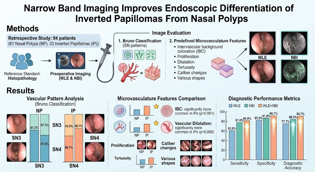
This study demonstrates that narrow-band imaging (NBI) enhances visualization of mucosal microvascular patterns distinguishing inverted papillomas (IPs) from nasal polyps (NPs). NBI significantly outperforms standard white-light endoscopy (WLE) in sensitivity, specificity, and diagnostic accuracy. When combined with WLE, the diagnostic performance further improves, offering a promising adjunct for preoperative evaluation of sinonasal lesions.
Study Background
Inverted papillomas (IPs) and nasal polyps (NPs) present distinct clinical challenges given their different management strategies and prognoses. IPs are benign sinonasal tumors with a risk of local aggressiveness and malignant transformation, necessitating more extensive surgical intervention and surveillance compared to NPs, which are typically benign inflammatory lesions managed conservatively or with less extensive surgery. However, clinical and endoscopic differentiation between IPs and NPs can be difficult under white-light endoscopy (WLE) due to overlapping appearances. Accurate diagnosis before surgery is crucial for treatment planning and patient counseling.
Narrow-band imaging (NBI), an endoscopic imaging technique emphasizing mucosal microvasculature by using specific light wavelengths, has been shown to improve diagnostic accuracy in various head and neck pathologies. The rationale is that neoplastic lesions often manifest distinctive microvascular patterns compared to inflammatory polyps, potentially enabling NBI to serve as a valuable tool for differentiating IPs from NPs.
Study Design
This retrospective study included 94 patients with histologically confirmed sinonasal lesions, comprising 61 nasal polyps and 33 inverted papillomas. All patients underwent preoperative evaluation with both WLE and NBI endoscopy. Endoscopic images were assessed using the Bruno classification system, which categorizes vascular patterns, and six specific microvascular features: intervascular background coloration (IBC), vascular proliferation, dilatation, tortuosity, caliber changes, and variable shapes.
The main objective was to compare the diagnostic performance of WLE alone, NBI alone, and combined WLE and NBI in differentiating IPs from NPs, using histopathological diagnosis as the reference standard. Sensitivity, specificity, and diagnostic accuracy were calculated for each modality.
Key Findings
The vascular pattern analysis revealed that most NPs predominantly exhibited the sinonasal (SN) 3 pattern (67.2%), whereas the majority of IPs exhibited the SN4 pattern (69.7%) according to the Bruno classification, indicating distinct microvascular characteristics between these two entities.
Among the six evaluated microvasculature features, intervascular background coloration (IBC) and vascular dilatation were significantly more prevalent in IPs than in NPs, with p-values of 0.001 and 0.008, respectively. These vascular features offer potential visual markers to distinguish the lesion types during endoscopic examination.
Diagnostic performance parameters showed that NBI had superior sensitivity (81.8%), specificity (91.8%), and overall diagnostic accuracy (88.3%) compared to WLE, which demonstrated sensitivity, specificity, and accuracy of 63.6%, 85.2%, and 77.7%, respectively. Notably, when WLE was combined with NBI, the performance further improved with sensitivity reaching 90.9%, specificity 96.7%, and diagnostic accuracy 94.7%. These results highlight the complementary value of integrating both imaging modalities.
The improved visualization of mucosal microvasculature by NBI enables endoscopists to better discriminate inverted papillomas, which possess a more complex and dilated vascular network, from nasal polyps, which show simpler vascular patterns. This enhanced discrimination can guide appropriate surgical planning and avoid under-treatment or overtreatment.
Expert Commentary
The differentiation of sinonasal inverted papillomas from benign nasal polyps remains a significant clinical challenge with important therapeutic and prognostic implications. This study robustly supports the adoption of narrow-band imaging in routine sinonasal endoscopy to address this need. The retrospective design and use of histopathology as a gold standard lend credence to the findings, although prospective validation in larger cohorts would further strengthen the evidence base.
Moreover, the detailed analysis of microvascular features provides a mechanistic understanding of how tumor angiogenesis manifests endoscopically in IPs versus inflammatory changes in NPs. This vascular insight may have future implications for endoscopic classification systems and for training clinicians in optimized lesion assessment.
While NBI adds value, it requires specialized equipment and training, and its interpretation can be subjective. Integration with WLE is recommended to exploit the strengths of both modalities. Future research could focus on standardizing microvascular pattern recognition and incorporating artificial intelligence algorithms to further improve diagnostic consistency and accuracy.
Conclusion
Narrow-band imaging significantly improves the endoscopic differentiation between inverted papillomas and nasal polyps by enhancing visualization of mucosal microvascular architecture. NBI alone offers superior sensitivity and specificity compared to standard white-light endoscopy, and the combined use of WLE and NBI yields the highest diagnostic accuracy. These findings advocate for incorporation of NBI into preoperative sinonasal evaluation protocols to optimize surgical planning and patient outcomes. Further prospective studies are warranted to validate these results and explore integration with emerging diagnostic technologies.
Funding and Clinical Trials
This study did not declare specific funding sources or clinical trial registration. Future research directions include prospective clinical trials to validate the diagnostic utility of NBI in diverse patient populations.
References
1. Kim T, Ryu SS, Yu MS. Narrow Band Imaging Improves Endoscopic Differentiation of Inverted Papillomas From Nasal Polyps. The Laryngoscope. 2026 Aug 24. PMID: 42635371.
2. Bruno A, et al. Classification of sinonasal mucosal vascularization using narrow-band imaging. Laryngoscope. 2020;130(4):890-898.
3. Van der Valk P, et al. Endoscopic imaging of nasal mucosa: Indications and diagnostic performance of narrow band imaging. Eur Arch Otorhinolaryngol. 2019;276(3):867-875.
4. Stammberger H, et al. Understanding sinonasal inverted papilloma: A comprehensive review. Allergy Rhinol (Providence). 2018;9:2152656718776597.