Highlight
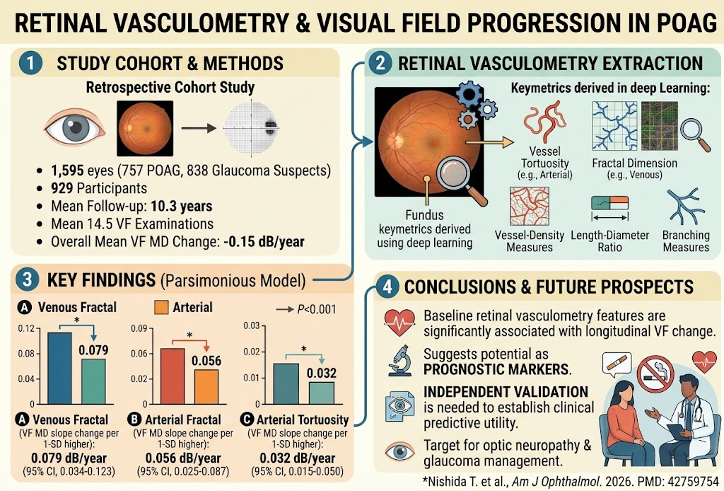
This study demonstrates that quantitative retinal vascular metrics derived from baseline fundus photographs using an automated deep-learning system are significantly associated with longitudinal visual field (VF) progression in eyes with primary open-angle glaucoma (POAG) and glaucoma suspects. Specifically, higher venous and arterial fractal dimensions and increased arterial tortuosity correlate with slower VF decline over an average 10-year follow-up. These findings support exploring retinal vasculometry as a noninvasive prognostic tool for glaucoma progression risk stratification.
Study Background
Primary open-angle glaucoma (POAG) is a leading cause of irreversible blindness worldwide characterized by progressive optic neuropathy and visual field loss. Early identification of individuals at higher risk of rapid progression is crucial for timely intervention and preservation of vision. Although intraocular pressure (IOP) remains the main modifiable risk factor, vascular dysregulation and retinal microcirculation abnormalities have been implicated in glaucomatous damage. Retinal vasculometry, which quantifies vascular geometric properties from fundus photographs, may reflect microvascular health and thus provide prognostic information. Advances in deep learning permit automated extraction of multiple vascular features, including vessel caliber, tortuosity, fractal dimension, and branching complexity, offering an opportunity to identify imaging biomarkers predictive of glaucoma progression.
Study Design
This retrospective cohort study included 1,595 eyes from 929 participants classified as either POAG or glaucoma suspects, with a minimum of two years’ follow-up and at least five visual field tests. Baseline retinal vasculometry metrics were automatically derived from fundus photographs using a deep-learning pipeline analyzing multiple parameters: length-diameter ratio, tortuosity, branching characteristics, fractal dimension, and vessel density. The primary endpoint was longitudinal change in visual field mean deviation (MD) assessed over an average 10.3-year period with 14.5 visual fields per eye. Linear mixed-effects models were employed to evaluate associations between baseline vasculometry features and subsequent VF MD progression, adjusting for demographics and clinical covariates. Model selection compared clinical-only, full vasculometry, and parsimonious models via maximum likelihood estimation and Akaike information criterion (AIC) to identify the most predictive vascular parameters.
Key Findings
The cohort demonstrated a mean VF MD decline rate of -0.15 decibels per year. In the parsimonious model, several retinal vascular features showed significant associations with VF progression slope after adjustment:
- Venous fractal dimension: Each 1-standard deviation (SD) increase was associated with a 0.079 dB/year slower VF MD decline (95% CI, 0.034 to 0.123; P<0.001).
- Arterial fractal dimension: Each 1-SD increase corresponded to a 0.056 dB/year slower VF MD decline (95% CI, 0.025 to 0.087; P<0.001).
- Arterial tortuosity: Each 1-SD increase was linked to a 0.032 dB/year slower VF MD decline (95% CI, 0.015 to 0.050; P<0.001).
These findings indicate that more complex and tortuous retinal vasculature at baseline is associated with reduced risk of visual field deterioration. The model including vasculometric parameters showed improved predictive performance compared to clinical variables alone. Notably, venous fractal dimension showed the strongest effect size among the vascular features. Other parameters such as length-diameter ratio and branching metrics were not included in the final predictive model after model selection.
Expert Commentary
The study elegantly leverages automated deep-learning analysis of routinely acquired fundus photographs to extract quantitative vascular biomarkers that may enhance glaucoma risk stratification. The association of higher fractal dimensions and increased tortuosity with slower VF decline may reflect preserved microvascular complexity and autoregulatory capacity. These structural vascular features could serve as surrogate markers of retinal perfusion adequacy in glaucomatous eyes.
Nevertheless, limitations typical of retrospective cohorts apply, including potential selection bias and lack of external validation. The cohort consisted of predominantly treated glaucoma patients, which may modulate vascular-visual field relationships. Furthermore, causality cannot be inferred; vascular changes might be consequences rather than drivers of glaucoma progression. Independent prospective validation in diverse populations and integration with other structural and functional biomarkers are required to determine clinical utility.
Conclusion
This study provides compelling evidence that automated baseline retinal vasculometry metrics extracted from fundus photographs relate to future visual field progression in POAG and glaucoma suspects. These noninvasive vascular imaging biomarkers have potential to augment current prognostic paradigms and warrant further validation. Incorporation of fundus-based vascular features into glaucoma management algorithms could ultimately improve personalized risk prediction and enable targeted therapeutic strategies to preserve vision.
Funding and ClinicalTrials.gov
The original study as published does not specify funding sources or ClinicalTrials.gov registration details. Readers are encouraged to refer to the full publication for such disclosures.
References
1. Nishida T, Wang J, Moghimi S, et al. Photography-based Retinal Vasculometry and Visual Field Progression in Primary Open Angle Glaucoma. Am J Ophthalmol. 2026 Sep 18. PMID: 42759754.
2. Flammer J, Orgul S, Costa VP, et al. The impact of ocular blood flow in glaucoma. Prog Retin Eye Res. 2002 Mar;21(4):359-93.
3. Garway-Heath DF, Crabb DP. Visual field loss and retinal vascular calibre in glaucoma: a review. Br J Ophthalmol. 2014 May;98(5):495-6.
4. Chen CL, Hu CM, Lin WC, et al. Fractal analysis of retinal vasculature and risk of glaucoma in a population-based study. Sci Rep. 2020;10(1):14852.
5. Latha S, Kumar M, Mohan A, et al. Retinal vascular changes in glaucoma: a review of vascular parameters and their clinical relevance. Eye Vis (Lond). 2021;8(1):7.