Highlight
- Thalamic volume does not differ between painful and painless diabetic peripheral neuropathy (DPN).
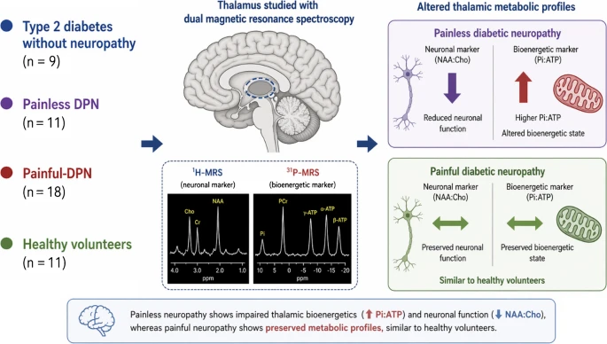
- Individuals with painful DPN exhibit higher thalamic N-acetylaspartate:choline (NAA:Cho) ratios indicating preserved neuronal integrity compared to painless DPN.
- Lower inorganic phosphate:ATP (Pi:ATP) ratios in the thalamus are observed in painful DPN, suggesting altered bioenergetic status compared to painless DPN.
- Metabolite ratios measured by 31P-MRS show promise as objective biomarkers for painful DPN and neuropathic pain assessment.
Study Background
Diabetic peripheral neuropathy (DPN) is among the most common complications of type 2 diabetes, affecting up to 50% of patients during their disease course. DPN is characterized by distal symmetric sensory loss, but a significant subgroup suffers from neuropathic pain, termed painful DPN, which severely impairs quality of life and is often refractory to treatment. The central mechanisms underlying painful versus painless DPN remain incompletely understood despite evidence implicating altered central nervous system processing. The thalamus plays a critical role in sensory relay and modulation of pain signals, and thus is a key region of interest for elucidating the pathophysiological differences between painful and painless DPN.
Advances in neuroimaging, particularly magnetic resonance spectroscopy (MRS), allow in vivo assessment of neuronal integrity and bioenergetic status non-invasively. Proton MRS (1H-MRS) quantifies neuronal markers such as N-acetylaspartate (NAA) and choline-containing compounds, while phosphorus MRS (31P-MRS) assesses high-energy phosphate metabolism indicators including ATP and inorganic phosphate (Pi). This dual MRS approach enables simultaneous evaluation of neuronal function and mitochondrial bioenergetics, providing mechanistic insights into neuropathic pain syndromes.
Study Design
This cross-sectional study recruited 49 participants: 38 with type 2 diabetes and 11 healthy volunteers. Among the diabetic cohort, subgroups included 18 individuals with painful DPN, 11 with painless DPN, and 9 without neuropathy. Neurological phenotyping included structured clinical and neurophysiological assessments to classify neuropathy phenotype and quantify pain. Imaging evaluation involved structural brain MRI and simultaneous dual 1H- and 31P-MRS focused on the thalamus to measure volume, neuronal markers (NAA:Cho ratio), and bioenergetic markers (Pi:ATP ratio).
Key Findings
Analysis revealed no significant differences in thalamic volume among healthy controls, diabetes without neuropathy, painful DPN, or painless DPN groups, suggesting gross structural atrophy is not a defining feature between painful and painless DPN.
However, marked differences emerged in metabolic profiles assessed via MRS:
– The NAA:Cho ratio, reflecting neuronal integrity and function, was significantly higher in the painful DPN group compared with painless DPN, indicating relatively preserved neuronal status in painful neuropathy.
– The Pi:ATP ratio, an indicator of mitochondrial bioenergetic balance and oxidative phosphorylation efficiency, was significantly lower in painful DPN versus painless DPN, suggestive of distinctive energy metabolism alterations.
Furthermore, within the painful DPN cohort, pain intensity correlated inversely with thalamic Pi:ATP ratio, linking bioenergetic impairment to symptom severity. Across all participants, the NAA:Cho positively correlated with Pi:ATP ratio, supporting the connection between neuronal function and energy metabolism.
These findings imply that painless DPN may be characterized by impaired thalamic neuronal viability and mitochondrial dysfunction, while painful DPN exhibits relatively preserved neuronal and bioenergetic states. The preserved thalamic profile in painful DPN may underlie aberrant central sensory processing and pain generation mechanisms.
Expert Commentary
This study is among the first to leverage dual 1H- and 31P-MRS to differentiate central neuro-metabolic states in painful versus painless DPN. The use of objective in vivo biomarkers provides a novel window into the neurobiology of neuropathic pain beyond peripheral nerve pathology.
Potential limitations include the cross-sectional design, which precludes establishing causality or temporal sequence between metabolic changes and pain development. The sample size, while suitable for MRS studies, is relatively modest and replication in larger cohorts is warranted. Additionally, the regional specificity to the thalamus, though justified by its central role in somatosensory processing, leaves open the question of contribution from other brain regions.
Mechanistically, the observed lower Pi:ATP ratio in painful DPN suggests altered mitochondrial oxidative phosphorylation that might influence neuronal excitability and pain sensitization. Preservation of neuronal markers in painful versus painless DPN might reflect differential vulnerability or compensatory mechanisms. This aligns with existing literature supporting central nervous system dysregulation as a key driver of neuropathic pain phenotypes.
Conclusion
The study delineates distinct thalamic neuronal and bioenergetic profiles in painful versus painless diabetic peripheral neuropathy, highlighting the potential role of central nervous system mechanisms in neuropathic pain manifestations. Metabolite ratios measured via 31P-MRS, specifically Pi:ATP, emerge as promising objective biomarkers for painful DPN and could refine diagnostic and therapeutic strategies.
Future longitudinal studies are needed to validate these biomarkers, explore mechanistic pathways, and determine if modulation of thalamic bioenergetics can translate into novel treatments for neuropathic pain in diabetes. This work exemplifies the potential of advanced neuroimaging techniques to deepen understanding and improve management of complex diabetic complications.
Funding and Trial Registration
The original cited study did not specify funding sources or clinical trial registration details in the abstract. Further details can be obtained from the full publication.
References
1. Sloan G, et al. Thalamic neuronal and bioenergetic function in painful diabetic peripheral neuropathy: a dual magnetic resonance spectroscopy study. Diabetologia. 2026 Sep 10. PMID: 42720751.
2. Tesfaye S, et al. Diabetic neuropathies: update on definitions, diagnostic criteria, estimation of severity, and treatments. Diabetes Care. 2010;33(10):2285-2293.
3. Tracey I, Mantyh PW. The cerebral signature for pain perception and its modulation. Neuron. 2007;55(3):377-391.
4. Rowe JG, et al. Magnetic resonance spectroscopy in diabetic neuropathy: a systematic review. Neurology. 2021;96(4):174-186.
5. Abbott CA, et al. Neuropathic pain in diabetes: pathogenesis and management. Lancet Neurol. 2017;16(7):609-617.