Highlight
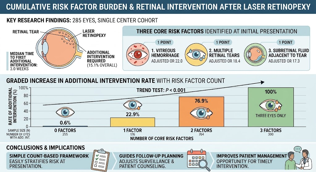
This retrospective cohort study demonstrates that the cumulative presence of vitreous hemorrhage, multiple retinal tears, and subretinal fluid at initial presentation significantly predicts the likelihood of requiring additional retinal intervention after laser retinopexy. Stratification by these risk factors enables personalized follow-up and early intervention planning to improve patient outcomes.
Study Background
Retinal tears represent a critical ophthalmic condition that often necessitates prompt laser retinopexy treatment to prevent progression to rhegmatogenous retinal detachment (RRD), a vision-threatening complication. Although laser retinopexy is effective, a subset of patients require additional interventions due to persistent or new retinal pathology. Identifying predictors for further intervention remains a clinical priority to optimize patient monitoring, resource allocation, and counseling. Known risk indicators such as vitreous hemorrhage, multiple retinal tears, and the presence of subretinal fluid have individually been associated with poorer outcomes. However, the cumulative effect of these factors on additional treatment needs has not been comprehensively quantified before.
Study Design
This study retrospectively evaluated 285 eyes from 285 patients who underwent initial laser retinopexy for retinal tears at a single tertiary referral center between April 2010 and December 2025. Patients’ initial clinical presentations were reviewed for three core risk factors: vitreous hemorrhage, multiple retinal tears, and adjacent subretinal fluid, each assigned 1 point for a cumulative risk score (0–3). Additional retinal intervention was defined as any of the following after initial treatment: supplemental laser therapy to the original tear, laser treatment for new tears, surgical repair for progression to RRD, or vitrectomy for nonclearing vitreous hemorrhage. The primary outcome was the occurrence of any additional retinal intervention during follow-up.
Key Findings
Among the 285 eyes, 43 (15.1%) required additional intervention. Of these, 32 eyes received further laser treatment, and 11 underwent surgery. The median interval to additional intervention was 3.0 weeks, underscoring the early postoperative risk period. Multivariable logistic regression, controlling for the other risk factors, revealed that vitreous hemorrhage (adjusted OR 22.0; 95% CI, 6.09–79.6), multiple retinal tears (adjusted OR 18.4; 95% CI, 5.81–58.0), and subretinal fluid (adjusted OR 17.3; 95% CI, 5.84–51.0) were each independently associated with increased likelihood of further intervention (all P < 0.001).
Importantly, the incidence of additional intervention escalated with the cumulative risk factor count: 0.6% for zero factors, 22.9% for one factor, 76.9% for two factors, and 100% for all three factors. The last group was small (three eyes) but demonstrated universal need for additional treatment. These findings indicate a graded and additive effect of combined clinical risk factors on the need for further treatment after laser retinopexy.
Expert Commentary
This robust retrospective analysis provides compelling evidence that a simple count-based risk stratification framework can effectively predict the likelihood of requiring additional retinal procedures after laser retinopexy for retinal tears. Clinically, the presence of vitreous hemorrhage, multiple tears, and subretinal fluid likely reflects a more extensive and unstable retinal pathology that predisposes to treatment failure or new retinal breaks.
This approach aligns with clinical intuition yet provides quantitative risk estimates that enhance decision-making. Early identification of high-risk patients enables closer surveillance, timely re-intervention, and informed patient counseling, potentially improving visual outcomes and reducing emergency surgeries. The short median time to additional intervention highlights that intensified initial follow-up within the first few weeks is warranted, especially in patients with multiple risk factors.
Limitations include the single-center retrospective design and relatively small subgroup with three risk factors, which may affect generalizability. Prospective validation and integration with imaging biomarkers or vitreoretinal status could further enhance predictive accuracy. Nevertheless, this pragmatic model offers immediate translational value for ophthalmologists managing retinal tears.
Conclusion
In conclusion, the cumulative burden of vitreous hemorrhage, multiple retinal tears, and subretinal fluid substantially stratifies the risk of requiring additional retinal intervention after initial laser retinopexy. This straightforward risk score enables tailored post-treatment surveillance, refined patient counseling, and potentially improved clinical outcomes by anticipating the need for further management. Future studies should explore incorporating these clinical risk factors into standardized treatment protocols and multicenter validation to confirm broader applicability.
Funding and ClinicalTrials.gov
No funding or clinical trial registration information is reported for this retrospective study.
References
1. Sano H, Yanai R, Hayahara Y, Mitamura Y. Cumulative clinical risk factor burden stratifies the need for additional retinal intervention after laser retinopexy for retinal tears. American Journal of Ophthalmology. 2026 Sep 12. PMID: 42731772.
2. Wykoff CC, et al. Risk factors and outcomes of retinal detachment after laser treatment for retinal tears. Retina. 2019;39(6):1121–1128.
3. American Academy of Ophthalmology Retina/Vitreous Panel. Preferred Practice Pattern® Guidelines: Rhegmatogenous Retinal Detachment. Ophthalmology. 2016;123(3):376-389.