Introduction and Context
Appendiceal abscess—an encapsulated collection arising from perforated appendicitis—is a frequent and clinically important variant of complicated appendicitis. Imaging is central to diagnosis, risk stratification and selection of management (conservative antibiotics, percutaneous drainage, or operative intervention). Yet, radiology reports and clinical pathways have long suffered from heterogeneous terminology (where precisely is the abscess?), varying severity descriptors, and inconsistent thresholds that determine whether to drain, operate, or observe. These inconsistencies hinder reproducibility, complicate research comparisons and can lead to variable outcomes.
In response, a multidisciplinary expert panel convened under the Italian Society of Research in Surgery and the Italian Society of Emergency and Trauma Surgery used a four-round modified Delphi process and an in-person conference (Rome, November 6, 2025) to produce a pragmatic, imaging-focused consensus. The resulting statements (Cirocchi et al., Surgery 2026) endorse standardized anatomotopographic localization, adoption of a reproducible radiological grading (the Jeffrey system), and structured imaging pathways tailored to adults, pregnant patients and follow-up after non-operative care.
Why this matters now: recent advances in cross-sectional imaging, wider use of percutaneous drainage, and evolving opinions about interval appendectomy demand harmonized language and pathways that map imaging findings to treatment choices. The consensus aims to reduce interpretative variability, improve decision-making, and enable meaningful multicenter research.
New Guideline Highlights
Key, practice-oriented takeaways from the 2025 Italian consensus:
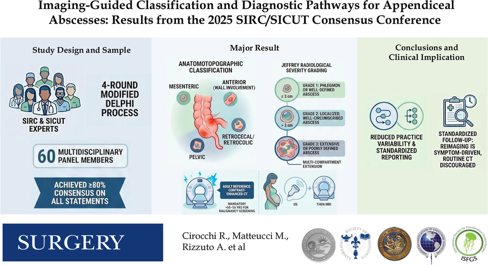
– Mandatory anatomotopographic classification. Every report of an appendiceal abscess should specify one of four locations: pelvic, mesenteric, retrocecal/retrocolic, or anterior with abdominal wall involvement.
– Adoption of the Jeffrey radiological grading (grades 1–3):
– Grade 1: phlegmon or small abscess ≤ 3 cm.
– Grade 2: well-circumscribed abscess > 3 cm.
– Grade 3: extensive or poorly-defined collection with multicompartment extension.
– Imaging pathways:
– Contrast-enhanced computed tomography (CECT) is the reference standard in adults.
– Selective CT use to evaluate for underlying malignancy—particularly in patients aged >50–55 years.
– Pregnancy: an ultrasound-first pathway, with MRI as second-line if ultrasound is inconclusive.
– Follow-up imaging after conservative management should be symptom-driven rather than routine, with selective ultrasound or CT for clinical deterioration or nonresolution.
– Structured reporting and standardized terminology are recommended to improve communication between radiologists and treating teams.
These statements achieved predefined consensus thresholds (≥80% for consensus; ≥95% for strong consensus on several items) and are designed to complement—not replace—clinical judgment.
Updated Recommendations and Key Changes from Prior Practice
What’s new or clarified compared with previous, less standardized approaches:
– From inconsistent descriptors to a required anatomical template. Previously, reports variably used terms such as “periappendiceal,” “pelvic,” or “deep right lower quadrant” without consistent mapping. The consensus mandates one of four anatomotopographic descriptors to improve reproducibility.
– Formal adoption of an explicit radiological grading (Jeffrey system). Prior practice often used vague terms (small vs large) or varying size cutoffs; the Jeffrey grading sets a clear 3-cm threshold between grade 1 and grade 2 and adds a grade for multicompartment disease.
– Pathway-driven imaging recommendations. While CT has long been used, the consensus explicitly endorses CECT as the adult reference, clarifies the age threshold for malignancy-focused CT evaluation, and formalizes pregnancy-specific ultrasound→MRI sequencing.
– Focused follow-up imaging guidance. Earlier practice varied widely between routine interval imaging and clinical follow-up. The consensus endorses symptom-driven follow-up, which may reduce unnecessary radiation and resource use.
Evidence driving these updates includes observational series correlating abscess size and complexity with drainage success and the growing body of data supporting ultrasound and MRI efficacy in pregnancy. The consensus also aligns with recent international emergency surgery guidance updates that emphasize individualized, imaging-informed care (see references).
Topic-by-Topic Recommendations
Below are the core recommendations framed for clinical use. Where possible, the consensus links imaging descriptors to practical management options.
Anatomotopographic classification (mandatory in reports):
– Pelvic: collection centered below the pelvic brim and abutting pelvic structures.
– Mesenteric: collection within the mesentery adjacent to the terminal ileum or cecum.
– Retrocecal/retrocolic: collection posterior to the cecum, potentially hidden from surface palpation.
– Anterior with abdominal wall involvement: collection extending to or involving the anterior abdominal wall or flank.
Clinical rationale: location informs risk of adjacent organ involvement, technical feasibility of percutaneous drainage and likelihood of symptom pattern (e.g., pelvic abscesses may present with urinary or pelvic symptoms).
Radiological severity grading (Jeffrey system):
– Grade 1: phlegmon or small abscess ≤ 3 cm. Recommended management: conservative antibiotics with clinical monitoring. Percutaneous drainage generally not indicated unless clinical deterioration occurs.
– Grade 2: well-circumscribed abscess > 3 cm. Recommended management: consider percutaneous image-guided drainage in addition to antibiotics; if drainage is not feasible, close clinical observation with antibiotics may be considered depending on patient stability and comorbidity.
– Grade 3: extensive/poorly defined collection with multicompartment extension. Recommended management: high likelihood of drainage requirement; early multidisciplinary (surgery + interventional radiology) planning; consider operative source control if percutaneous drainage is not feasible or if clinical deterioration ensues.
Practical size thresholds: the 3-cm cutoff reflects pooled observational data suggesting low likelihood of successful drainage below that size and higher success/need-to-drain above it. Clinical context (e.g., persistent fever, sepsis, immunosuppression) may override size-based guidance.
Imaging modality recommendations:
– Adults: contrast-enhanced CT with portal venous phase is the reference imaging modality because it reliably defines abscess extent, relationships to bowel and other organs, and suitability for percutaneous access.
– Older adults (>50–55 years): consider CT with attention to ruling out an underlying neoplasm—cancer presenting as complicated appendicitis or appendicular abscess becomes more likely with age.
– Pregnancy: ultrasound as first-line; if ultrasound is inconclusive and clinical suspicion remains, non-contrast MRI (or MRI without gadolinium) is recommended. CT should be used only when MRI is unavailable and clinical risk justifies radiation exposure.
– Children: ultrasound-first approach; low-dose CT reserved for equivocal US or when precise anatomic delineation is necessary for drainage planning.
Structured reporting elements (minimum):
– Anatomotopographic classification (pelvic/mesenteric/retrocecal/ anterior with abdominal wall involvement).
– Size(s) of abscess (maximum diameter) and volume estimate if possible.
– Jeffrey grade (1–3) assigned.
– Presence of fistula, free intraperitoneal air, or generalized peritonitis.
– Relationship to adjacent organs or bowel (for drainage planning).
– Presence of suspicious lesions suggesting underlying malignancy (age-tailored comment).
Follow-up imaging and clinical monitoring:
– Routine interval imaging is not recommended after conservative management.
– Symptom-driven imaging is advised: re-image only if clinical deterioration, persistent fever, rising inflammatory markers, or failure to improve within an expected timeframe.
– For patients who undergo percutaneous drainage, cross-sectional imaging may be used to confirm drain position or detect residual collections if clinical response is inadequate.
Special populations:
– Pregnancy: ultrasound-first, MRI-second. Involvement of obstetrics and maternal–fetal medicine for shared decision-making.
– Elderly and immunocompromised patients: lower thresholds for CT and for drainage due to higher risk of deterioration.
– Pediatric patients: ultrasound-first, use low-dose CT selectively; coordinate with pediatric surgery.
Interval appendectomy and long-term considerations:
– The consensus does not mandate routine interval appendectomy after successful non-operative management. Decisions should be individualized based on recurrence risk, patient preferences, diagnostic findings (e.g., suspicious lesions on CT), and local resources.
Expert Commentary and Insights
The expert panel emphasized that standardization improves both clinical care and research. Key points from the panel’s discussion included:
– Language matters: anatomotopographic descriptors reduce ambiguity. A pelvic abscess and a retrocecal collection are clinically different.
– Size matters, but context matters more. The Jeffrey 3-cm threshold is a practical dividing line, but comorbidity, sepsis, immunosuppression and technical feasibility influence the decision to drain.
– Imaging should drive, not dictate, management. The consensus repeatedly states that clinical judgment and multidisciplinary discussion remain critical—particularly for grade 2 lesions near the 3-cm cutoff and for grade 3 multicompartment disease.
– Pregnancy requires special pathways: pregnant patients benefit from ultrasound and MRI-first strategies. Radiologists and surgeons must coordinate care with obstetrics.
Areas of controversy and future research needs:
– The necessity and timing of interval appendectomy remain debated. The panel acknowledged heterogeneous international practice and recommended individualized decision-making while calling for prospective data.
– Optimal thresholds for drainage and the comparative effectiveness of early appendectomy versus percutaneous drainage remain areas ripe for multicenter prospective trials.
– Validation of the anatomotopographic–Jeffrey integrated approach in prospective registries is needed to confirm predictive value for drainage success, recurrence risk and clinical outcomes.
Practical Implications for Clinicians
Operational takeaways to implement the consensus in daily practice:
– Radiology departments should adopt structured report templates that include the four-location anatomotopographic descriptor and the Jeffrey grade.
– Emergency departments and surgical teams should use the grade-location framework to guide early management decisions and to communicate clearly with interventional radiology.
– Consider a multidisciplinary protocol: adult patients with grade 2–3 abscesses should have combined surgical and interventional radiology planning available to streamline timely drainage when indicated.
– For patients >50–55 years, use CT to rule out neoplasm as part of the diagnostic workup for appendiceal abscess.
– Educate trainees and frontline clinicians on the standardized terminology to ensure consistent documentation and to facilitate enrollment in prospective studies.
Patient vignette (illustrative):
– John, a 52-year-old man, presents with 5 days of right lower quadrant pain and fever. CT (CECT) shows a well-circumscribed 4.5-cm collection located in the mesentery adjacent to the cecum (Jeffrey grade 2). Because of his age and abscess size, the team arranges CT-guided percutaneous drainage plus IV antibiotics. The structured report documents “mesenteric” location and Jeffrey grade 2 and highlights no radiological signs suggestive of malignancy. The drain produces purulent fluid and John improves rapidly.
This vignette illustrates how clear imaging descriptors (location + grade + size) speed decision-making and guide appropriate interventional therapy.
References
– Cirocchi R, Matteucci M, Rizzuto A, et al. Imaging-guided classification and diagnostic pathways for appendiceal abscesses: Results from the 2025 Italian Society of Research in Surgery/Italian Society of Emergency and Trauma Surgery consensus conference. Surgery. 2026 Jun 6;197:110383. PMID: 42378837. https://pubmed.ncbi.nlm.nih.gov/42378837/
– Di Saverio S, Podda M, De Simone B, et al. Diagnosis and treatment of acute appendicitis: 2020 update of the WSES Jerusalem guidelines. World J Emerg Surg. 2020;15:27. doi:10.1186/s13017-020-00306-3.
– American College of Radiology. ACR Appropriateness Criteria® Right Lower Quadrant Pain—Suspected Appendicitis. ACR.org (latest revision online). (Access the ACR Appropriateness Criteria at www.acr.org)
– Mentula P, Leppäniemi A. Management of complicated intra-abdominal infections in adults: current recommendations of the World Society of Emergency Surgery. J Wound Care. (Contextual sources on percutaneous drainage and conservative management outcomes.)
Note: The consensus complements extant international guidance (including recent WSES updates) and is intended to be jointly applied with clinical judgment. The group recommends prospective multicenter validation and registries to test the prognostic performance of the anatomotopographic–Jeffrey integrated framework.
Closing
The 2025 Italian consensus offers a pragmatic, imaging-led approach that can be implemented immediately: mandatory anatomotopographic localization, adoption of the Jeffrey radiological grading, and pathway-based imaging advice tailored to adults, older patients, pregnant patients and children. Standardized reporting and clear ties between imaging findings and management options promise to reduce variability, clarify communication and create a dependable common language for clinicians and researchers. Prospective validation will determine whether this harmonized approach improves patient-centered outcomes—such as drainage success, avoidance of unnecessary surgery, recurrence rates and overall safety.