Highlight
- Local regrowth after neoadjuvant chemoradiotherapy (nCRT) in esophageal cancer primarily involves the mucosal and submucosal layers.
- Most regrowth cases encompass multiple esophageal wall layers, with persistent patterns over time.
- Endoscopy is supported as a crucial tool for detecting regrowth due to its accessibility to mucosal and submucosal lesions.
- Despite local control potential, nodal involvement poses a challenge; thus, surgery remains necessary for confirmed regrowth.
Study Background
Esophageal cancer remains a challenging malignancy with high morbidity and mortality, particularly in locally advanced stages. Neoadjuvant chemoradiotherapy (nCRT) followed by surgery constitutes a standard treatment approach aimed at tumor downsizing and improving resectability. However, concerns about overtreatment and surgery-related morbidity have stimulated interest in active surveillance strategies for patients achieving clinical complete response (cCR) to nCRT. In this context, timely and accurate detection of tumor regrowth is critical to optimize patient outcomes. Prior to the SANO trial, evidence on regrowth patterns post-nCRT was limited, which hindered the refinement of surveillance protocols and complicated decisions about potential endoscopic interventions versus surgical management.
Study Design
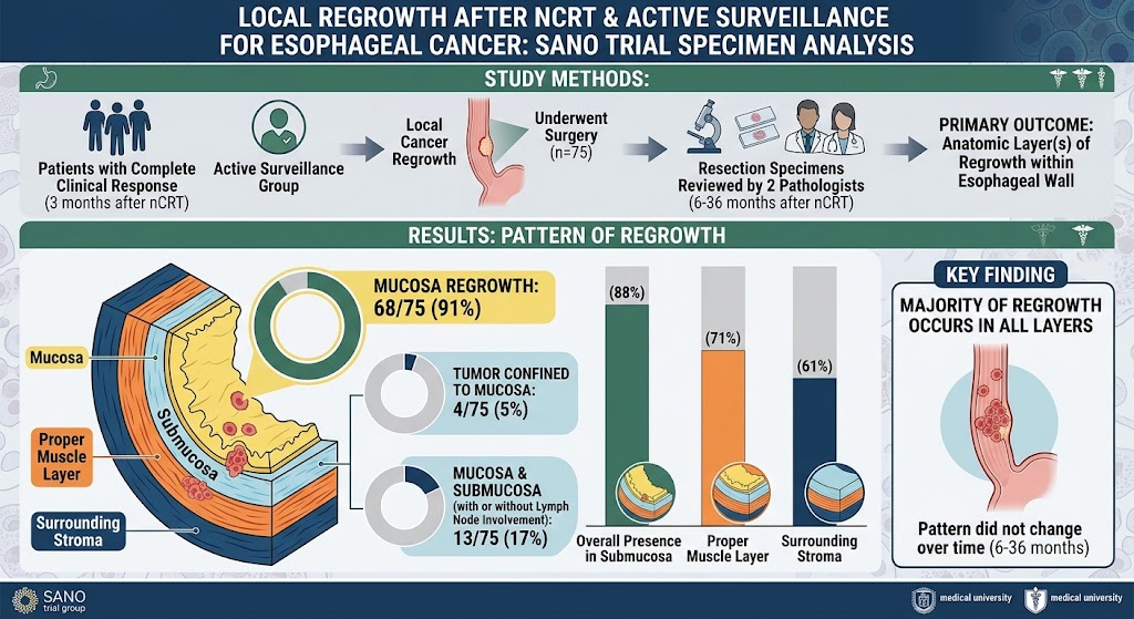
The investigation was a retrospective cohort analysis nested within the SANO trial framework. It included patients who initially achieved a complete clinical response three months post-nCRT and entered an active surveillance protocol. Among this cohort, individuals exhibiting local tumor regrowth who subsequently underwent surgical resection were selected for detailed pathological examination. Two expert pathologists independently reviewed 75 available esophagectomy specimens obtained between 6 and 36 months after nCRT. The primary aim was to determine regrowth localization within the esophageal wall’s histological layers, including mucosa, submucosa, proper muscle layer, surrounding stroma, and lymph nodes.
Key Findings
The pathological evaluation revealed that cancer cells were present in the mucosal layer in 91% (68/75) of cases. Notably, only 5% (4/75) of patients demonstrated tumor confined strictly to the mucosa. The mucosa and submucosa both harbored tumor in 17% (13/75), among which 4 patients also had lymph node metastases. Extending deeper, the submucosa involved tumor presence in 88%, the proper muscle layer in 71%, and the surrounding stromal tissue in 61% of specimens. These data clearly indicate that regrowth typically occurs across multiple contiguous histologic layers rather than isolated to the mucosa. Importantly, the distribution pattern of regrowth did not fluctuate significantly over the 6 to 36 month surveillance period, suggesting a stable biological behavior once regrowth initiates.
Clinically, these findings have important implications. Detection efforts should prioritize mucosal and submucosal evaluation using high-resolution endoscopy because the majority of local recurrences begin or are present at these superficial layers. The persistence of regrowth patterns across time underscores the relevance of regular surveillance intervals to timely detect recurrence. The high frequency of deeper invasion and nodal involvement also highlights the limitations of current clinical staging modalities post-nCRT, which may underestimate subclinical nodal disease.
Expert Commentary
Experts acknowledge this work as a valuable contribution toward understanding esophageal cancer dynamics after nCRT. Dr. Steven G.S.G. Gangaram et al. emphasize that while endoscopic techniques can effectively detect mucosal and submucosal regrowth, the risk of accompanying lymph node metastases justifies continued reliance on surgical resection for definitive management of detected regrowth. This aligns with current guidelines, which recommend surgery in patients with confirmed recurrence despite an initial strategy of active surveillance.
Limitations include the retrospective nature and potential selection biases of patients undergoing salvage surgery, possibly reflecting a subset with more clinically apparent or symptomatic regrowth. Moreover, innovations in imaging or molecular markers could improve nodal staging and reduce surgical overtreatment in the future. The study’s consistent pathological methodology and multi-layer analysis strengthen its reliability.
Conclusion
The analysis of surgical specimens from the SANO trial cohort confirms that local tumor regrowth after nCRT for esophageal cancer predominantly involves the mucosa and submucosa layers, accompanied frequently by deeper wall and nodal infiltration. These findings validate endoscopy as an essential tool in surveillance protocols to detect local regrowth early. However, given the risk of occult nodal disease and limitations in current staging accuracy, surgery remains the cornerstone for managing confirmed regrowth. Future research should focus on improving non-invasive detection of nodal involvement and potentially refining criteria for less invasive interventions.
Funding and ClinicalTrials.gov
The SANO trial is a multicenter study supported by academic institutions and clinical research grants. Details on trial registration and funding sources can be found in the primary publication (PMID: 42625251).
References
1. Gangaram Panday SSG, Oudijk L, Lagarde SM, et al. Local Regrowth After Neoadjuvant Chemoradiotherapy and Active Surveillance for Esophageal Cancer: An Analysis of Surgical Specimens From the SANO Trial. Ann Surg. 2026 Aug 21. PMID: 42625251.
2. van der Wilk BJ, et al. Active Surveillance in Clinically Complete Responders After Neoadjuvant Chemoradiotherapy for Esophageal Cancer. J Thorac Oncol. 2020;15(3):498-505.
3. Shapiro J, et al. Neoadjuvant Chemoradiotherapy Plus Surgery Versus Surgery Alone for Esophageal or Junctional Cancer (CROSS): Long-term Results of a Randomized Controlled Trial. Lancet Oncol. 2015;16(9):1090-1098.