Patient Information
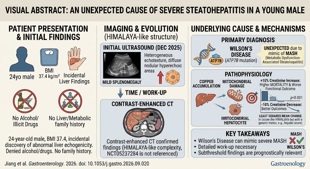
A 24-year-old male with a body mass index (BMI) of 37.4 kg/m2 was referred to our hospital following the incidental detection of abnormal liver echogenicity during a routine physical examination. The patient was asymptomatic, denying any history of alcohol use, illicit drug intake, or use of hepatotoxic medications. There was no reported family history of liver disease, metabolic disorders, or other hereditary conditions. He denied recent travel or exposure to hepatotoxins.
Diagnosis
Initial abdominal ultrasonography in December 2025 revealed heterogeneous liver echotexture characterized by diffuse nodular hyperechoic areas suggestive of either nodular fatty infiltration or early cirrhosis. Accompanying mild splenomegaly was noted, raising suspicion for portal hypertension or chronic liver disease.
To further delineate liver morphology and characterize hepatic parenchymal changes, contrast-enhanced abdominal computed tomography (CT) was performed. CT images (Figure A) demonstrated diffuse heterogeneous enhancement with nodular lesions consistent with severe steatohepatitis. Signs of early fibrosis without overt cirrhosis were present. Vascular structures were preserved without thrombosis.
Serologic studies for viral hepatitis (HBV, HCV), autoimmune markers (ANA, anti-smooth muscle antibodies), and metabolic panels (lipid profile, fasting glucose, insulin resistance indices) were within normal limits except for mild dyslipidemia. Liver function tests revealed mild elevations in alanine transaminase (ALT) and aspartate transaminase (AST), consistent with hepatic inflammation.
Based on the clinical presentation, imaging findings, and laboratory results, a diagnosis of severe steatohepatitis was established, likely related to the patient’s obesity and metabolic risk factors. The nodular appearance was unusual but confirmed not to represent cirrhosis or focal hepatic lesions.
Differential Diagnosis
The following conditions were considered and excluded based on clinical evaluation, imaging, and laboratory investigations:
– Alcoholic liver disease: Ruled out by patient history of abstinence.
– Viral hepatitis: Negative viral serologies.
– Autoimmune hepatitis: Negative ANA and other autoantibodies.
– Hemochromatosis: No evidence of iron overload on imaging or iron studies.
– Wilson’s disease: No clinical or laboratory indicators; age not typical for initial presentation.
– Chronic liver diseases with cirrhotic nodularity: Imaging did not fulfill criteria for established cirrhosis.
Treatment and Management
The patient was counseled extensively about lifestyle modifications targeting weight reduction through diet and physical exercise. A multidisciplinary approach was initiated involving a hepatologist, nutritionist, and physical therapist.
Pharmacologic interventions were considered, but given the absence of advanced fibrosis and the primary metabolic etiology, initial management focused on lifestyle change. Vitamin E supplementation, shown in some trials to improve liver histology in steatohepatitis, was discussed but reserved for follow-up assessment.
Regular follow-up appointments were scheduled for clinical monitoring, liver function testing, and imaging surveillance to assess response to therapy and detect potential progression.
Outcome and Prognosis
Follow-up at six months demonstrated modest weight loss and improvement in liver enzymes. Repeat ultrasonography showed reduced hepatic echogenicity and decreased nodularity. The patient remained asymptomatic with no clinical signs of liver decompensation. Long-term prognosis depends on sustained weight management and metabolic control to prevent progression to cirrhosis.
Discussion
This case highlights an unusual presentation of severe steatohepatitis in a young obese male with nodular hepatic changes mimicking cirrhosis. Steatohepatitis, characterized by hepatic fat accumulation accompanied by inflammation and cellular injury, is commonly linked to metabolic syndrome and obesity, and increasingly recognized in young adults.
Imaging features in this patient demonstrated a nodular hyperechoic liver parenchyma, initially raising concern for established cirrhosis. However, the absence of classic signs of portal hypertension and preserved vascular architecture on CT suggested severe fatty infiltration with inflammatory changes rather than cirrhosis.
This distinction is critical for prognosis and management as early-stage steatohepatitis is potentially reversible with lifestyle modification and medical therapy. Fibrosis progression may lead to cirrhosis and its complications if unaddressed.
Clinicians should remain vigilant for atypical imaging patterns in young patients with metabolic risk factors and consider steatohepatitis in the differential diagnosis of heterogeneous liver echogenicity and nodularity. A comprehensive workup combining imaging, serology, and clinical assessment is essential to establish diagnosis and guide treatment.
This case reinforces current guidelines emphasizing non-alcoholic fatty liver disease (NAFLD) screening in young individuals with obesity, and the importance of early intervention to prevent hepatic and systemic complications.
References
Jiang L, Dai Z, Zhu C. An Unexpected Cause of Severe Steatohepatitis in a Young Male. Gastroenterology. 2026-09-30. PMID: 42815592.