Highlight
Emergency medical services (EMS) frequently activate STEMI pathways based on prehospital suspicion before confirmatory testing. Among patients with paramedic-suspected STEMI, only about 60% received confirmed STEMI diagnoses at discharge, highlighting diagnostic heterogeneity. Despite this, rapid access to reperfusion therapies remains critical, yet early diagnostic refinement for alternate serious conditions is equally important to optimize care and outcomes.
Study Background
ST-segment elevation myocardial infarction (STEMI) is a life-threatening cardiac emergency requiring timely reperfusion to reduce morbidity and mortality. Prehospital identification of STEMI by paramedics triggers emergency pathways designed to expedite patient transfer and initiation of reperfusion strategies such as percutaneous coronary intervention (PCI). However, EMS activation relies on clinical assessment and prehospital electrocardiography (ECG) before confirmatory biomarker or angiographic evidence is available, leading to diagnostic uncertainty. Understanding the spectrum of final diagnoses, interventions, and outcomes after paramedic suspicion of STEMI is essential for optimizing STEMI care pathways and resource allocation.
Study Design
This retrospective cohort study utilized data from the Victorian Ambulance STEMI Quality Initiative registry, linked with hospital follow-up, analyzing cases from January 2020 to June 2025. Included were patients for whom paramedics suspected STEMI based on prehospital evaluation and ECG findings, activating STEMI protocols. Discharge diagnoses were classified using ICD-10-AM codes into five categories: STEMI; non-ST-elevation acute coronary syndrome (NSTE-ACS) or other coronary syndromes; other cardiac diagnoses; noncardiac diagnoses; and symptom-based diagnoses without a specific cardiac or noncardiac cause. Key endpoints included out-of-hospital management, rates of coronary angiography, percutaneous coronary intervention, complications, length of hospital stay, inhospital and 30-day mortality, and longer-term mortality (median follow-up 2.8 years). Multivariable logistic and Cox regression models adjusted for demographic and clinical covariates estimated associations with outcomes, reported as odds ratios (OR) or hazard ratios (HR) and 95% confidence intervals (CI).
Key Findings
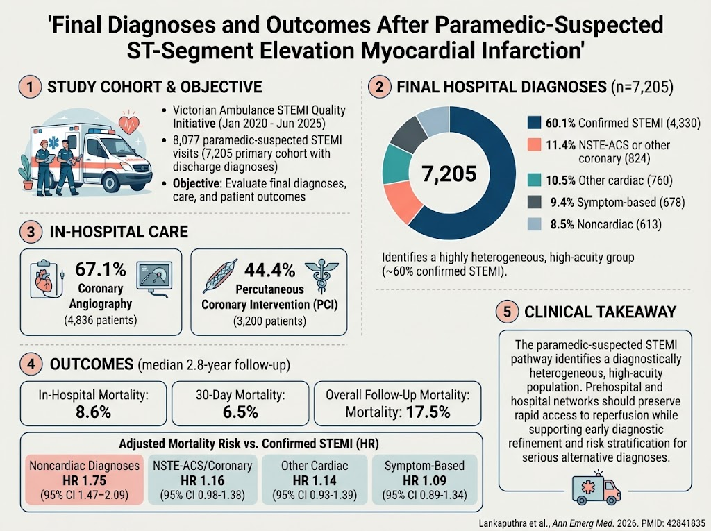
Of 8,077 paramedic-suspected STEMI cases with hospital follow-up data, 7,205 (89.2%) had a recorded discharge diagnosis forming the main analysis cohort. Within this group:
- 4,330 patients (60.1%) were confirmed to have STEMI.
- 824 (11.4%) had NSTE-ACS or other coronary syndromes.
- 760 (10.5%) had other cardiac diagnoses (e.g., arrhythmias, myocarditis).
- 613 (8.5%) had noncardiac diagnoses (e.g., pulmonary embolism, gastrointestinal causes).
- 678 (9.4%) had symptom-based diagnoses without a definitive etiology.
Coronary angiography was performed in 4,836 (67.1%) patients, and PCI in 3,200 (44.4%). Inhospital mortality was 8.6%, 30-day mortality was 6.5%, and mortality during median 2.8 years follow-up was 17.5%. After adjusting for confounders, mortality during follow-up was significantly higher among patients with noncardiac diagnoses compared with confirmed STEMI (HR 1.75, 95% CI 1.47 to 2.09). Mortality for other cardiac diagnoses (HR 1.14, 95% CI 0.93 to 1.39), NSTE-ACS or other coronary syndromes (HR 1.16, 95% CI 0.98 to 1.38), and symptom-based diagnoses (HR 1.09, 95% CI 0.89 to 1.34) was not materially different from confirmed STEMI.
Notably, 872 (10.8%) cases lacked a recorded discharge diagnosis, which may represent incomplete documentation, transfers, or early mortality.
Expert Commentary
This study provides robust, real-world evidence on the heterogeneity of final diagnoses after paramedic-suspected STEMI and their associated outcomes in a large, statewide cohort. The finding that only 60% of suspected STEMI patients have confirmed STEMI at discharge reflects both the challenges and necessary caution of prehospital STEMI identification. Over-triage is balanced by the critical imperative of timely reperfusion, which improves prognosis in true STEMI cases.
The elevated long-term mortality in patients with noncardiac diagnoses emphasizes the need for thorough early diagnostic evaluation within the STEMI pathway. Alternative serious conditions such as pulmonary embolism or aortic dissection may present with chest pain and ECG changes, warranting comprehensive risk stratification and tailored management.
Limitations of this registry study include potential misclassification bias, lack of information on biomarker specifics and timing, and absence of details regarding reasons for absence of discharge diagnosis. Generalizability is limited to similar prehospital settings and should not be extrapolated to all chest pain presentations or confirmed STEMI populations.
Conclusion
Paramedic-suspected STEMI captures a diagnostically diverse and clinically high-acuity cohort, with approximately 60% confirmed STEMI after hospital evaluation. Maintaining emergency pathway efficiency to ensure rapid reperfusion remains paramount, but integration of early diagnostic refinement and risk stratification processes is essential to optimize patient outcomes and resource use. Future strategies should focus on enhanced prehospital diagnostics and decision support to balance sensitivity and specificity in STEMI identification.
Funding and Registration
The study was conducted under the Victorian Ambulance STEMI Quality Initiative registry. No specific funding was reported. Clinical trial registration information was not provided.
References
1. Lankaputhra M, Bishop A, Wijaya J, et al. Final Diagnoses and Outcomes After Paramedic-Suspected ST-Segment Elevation Myocardial Infarction. Ann Emerg Med. 2026 Oct 6;PMID: 42841835.
2. O’Gara PT, Kushner FG, Ascheim DD, et al. 2013 ACCF/AHA guideline for the management of ST-elevation myocardial infarction. J Am Coll Cardiol. 2013;61(4):e78-140.
3. Ibanez B, James S, Agewall S, et al. 2017 ESC Guidelines for the management of acute myocardial infarction in patients presenting with ST-segment elevation. Eur Heart J. 2018;39(2):119-177.