Highlight
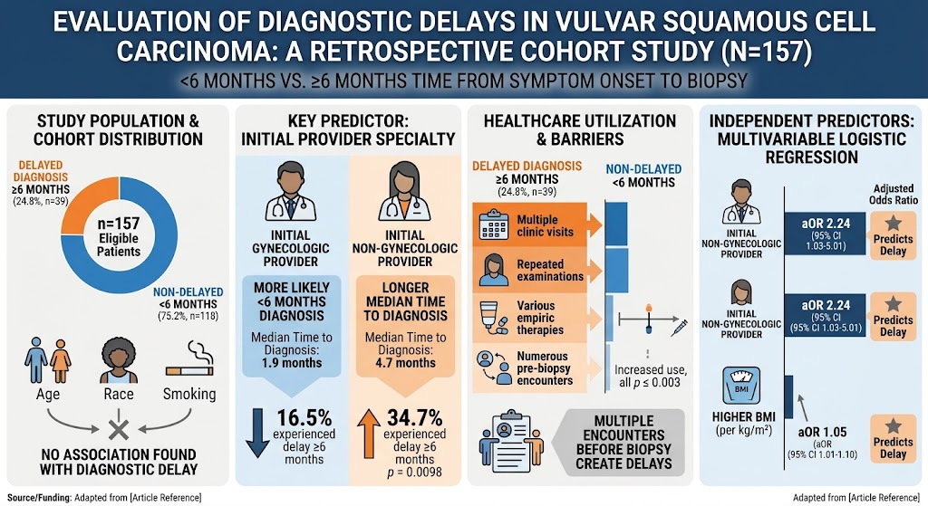
- Nearly 25% of vulvar squamous cell carcinoma (SCC) patients experience diagnostic delays of 6 months or more.
- Diagnostic delays are strongly linked to initial evaluation by non-gynecologic providers and higher patient BMI rather than tumor characteristics or socioeconomic factors.
- Patients with delayed diagnosis undergo significantly more clinic visits, empirical treatments, and medical encounters before biopsy.
- Improving early biopsy practices and timely gynecologic referrals could reduce diagnostic delays and improve outcomes in vulvar SCC.
Study Background
Vulvar squamous cell carcinoma (SCC), though relatively rare, represents a serious gynecologic malignancy with significant morbidity and mortality when diagnosis is delayed. Early diagnosis is critical because vulvar lesions can be subtle, and symptoms are often nonspecific, leading to frequent misdiagnosis or delayed clinical suspicion. Diagnostic delays contribute to disease progression, complicate treatment, and negatively impact progression-free and overall survival. While patient-level factors such as demographics have been posited to influence diagnosis timing, the contribution of provider and healthcare system factors remains less elucidated. This study aims to clarify factors associated with delayed diagnosis to inform targeted interventions to expedite identification and treatment of vulvar SCC.
Study Design
This retrospective cohort study was conducted at a single institution, including 157 patients diagnosed with vulvar SCC between 2009 and 2020. The patients were categorized based on time from symptom onset to biopsy, using a threshold of less than 6 months (<6 months) versus 6 months or more (≥6 months) to define delayed diagnosis. The analysis collected demographic data (age, race, smoking status, body mass index), tumor characteristics (size, stage), provider specialty at initial evaluation, insurance and income data, and healthcare utilization metrics including the number of clinical visits and treatments before biopsy. The study also examined progression-free survival (PFS) and overall survival (OS) outcomes.
Key Findings
Of the 157 women included, 39 (24.8%) experienced a delay of 6 months or longer before biopsy-confirmed diagnosis. Interestingly, demographic and tumor-specific factors such as age, race, smoking status, presence of dermatologic conditions, tumor size, stage at diagnosis, insurance status, and income level did not significantly differ between patients with timely and delayed diagnosis.
Significant differences emerged in healthcare interactions: patients with delayed diagnosis had a higher median number of clinic visits, examinations, empirical therapies, and overall healthcare encounters prior to biopsy (all with p ≤ 0.003). This suggests a protracted diagnostic process rather than straightforward clinical recognition.
Critically, the initial provider specialty was a strong determinant. Patients first seen by gynecologic providers had a median time to diagnosis of 1.9 months, whereas those initially evaluated by non-gynecologic providers faced a significantly longer median delay of 4.7 months (p < 0.0001). Moreover, 34.7% of patients initially evaluated by non-gynecologic providers experienced diagnostic delays of 6 months or longer compared to 16.5% for those presenting first to gynecologic providers (p = 0.0098).
Multivariable logistic regression identified two independent predictors of delayed diagnosis: higher body mass index (BMI) (adjusted odds ratio [aOR], 1.05 per unit increase; 95% confidence interval [CI], 1.01-1.10) and initial evaluation by a non-gynecologic provider (aOR, 2.24; 95% CI, 1.03-5.01). These findings highlight that delays are strongly influenced by provider familiarity and possibly patient anatomical factors complicating exam.
Although the study examined survival outcomes, detailed PFS and OS comparisons based on diagnostic delay were not explicitly reported in the abstract.
Expert Commentary
The study underscores a critical gap in vulvar cancer care: diagnostic delays are less about patient demographics or tumor presentation and more about healthcare provider awareness, clinical suspicion, and system navigation. Gynecologic providers, given their specialized training, appear more adept at timely identification and biopsy of suspicious vulvar lesions. In contrast, non-gynecologic clinicians—who often represent initial points of contact, including primary care, dermatology, or other specialties—may lack sufficient awareness or confidence to promptly biopsy vulvar lesions, resulting in prolonged diagnostic intervals.
Higher BMI as a predictor of delay may relate to examination challenges or implicit biases that may delay thorough vulvar evaluation. This finding calls for increased vigilance and possibly tailored approaches in the examination of obese patients.
Limitations include the single-institution retrospective design, which may limit broader generalizability. Additionally, socioeconomic factors, while analyzed, might require further granular investigation. The study did not differentiate delays attributable to patient versus system factors within the healthcare visit sequence.
Evidence from guidelines and expert opinion consistently advocates for early biopsy of persistent vulvar lesions and the importance of specialized referral pathways. This study provides robust data to support educational interventions aimed at non-gynecologic providers to improve early suspicion and sampling of potential vulvar malignancies.
Conclusion
Delayed diagnosis of vulvar squamous cell carcinoma is a substantial barrier to optimal patient outcomes. This delay is predominantly influenced by provider-related factors, especially initial evaluation by non-gynecologic clinicians, and patient factors such as elevated BMI that may complicate clinical assessment. Prompt biopsy of suspicious lesions, enhanced education for non-gynecologic providers, and streamlined referral to gynecologic specialists are critical strategies to reduce diagnostic delays. These interventions hold promise to improve survival and quality of life for women with vulvar SCC, emphasizing the urgent need to raise awareness and improve diagnostic pathways in vulvar cancer care.
Funding and ClinicalTrials.gov
The original study was institutionally approved with IRB oversight; details on funding sources or clinical trial registration were not provided in the report.
References
1. Sakach JC, Morton M, Velasquez JM, et al. No time to waste: Evaluation of diagnostic delays in vulvar cancer. Gynecol Oncol. 2026 Aug 3;212:43-49. PMID: 42546435.
2. Hinten F, van der Avoort IA, Massuger LF, et al. Delay in the diagnosis of vulvar cancer: An important determinant of prognosis. Gynecol Oncol. 2016;140(2):266-270.
3. National Comprehensive Cancer Network. Vulvar Cancer (Version 2.2023). https://www.nccn.org/professionals/physician_gls/pdf/vulvar.pdf
4. van de Nieuwenhof HP, de Hullu JA, Hollema H, et al. Carcinoma of the vulva: Problems in early diagnosis. Eur J Gynaecol Oncol. 2008;29(6):598-601.
(Note: The above references include the original study and relevant guideline and studies to contextualize findings.)