Introduction and Context
The American Society of Breast Surgeons (ASBrS), in collaboration with the Society of Breast Imaging (SBI) and the College of American Pathology (CAP), released a joint guideline in 2026 addressing the increasingly common clinical problem of proliferative breast lesions with atypia and lobular carcinoma in situ (LCIS). The guideline (Nakhlis et al., JAMA Surgery 2026) responds to persistent uncertainties about which core-biopsy–detected atypical lesions require surgical excision, which can be safely observed, how to apply chemoprevention, and when pathology second opinions are necessary. The recommendations are intended to standardize care, reduce unnecessary surgery, and ensure that patients at elevated future breast cancer risk receive appropriate counseling and preventive options.
Why this guidance matters now
– The use of image-guided core needle biopsy has grown, increasing detection of focal atypia (flat epithelial atypia [FEA], atypical ductal hyperplasia [ADH], atypical lobular hyperplasia [ALH]) and LCIS variants (classic LCIS [C‑LCIS], pleomorphic LCIS [P‑LCIS], and florid LCIS [F‑LCIS]).
– Historical variability in practice has led to both over-treatment (unnecessary excisions) and under-treatment (missed upgrade to carcinoma).
– New data on upgrade rates, long-term risk, and the effectiveness of chemoprevention prompted a multispecialty reassessment to align surgical, radiologic, and pathologic approaches.
Primary citation
Nakhlis F, Bedrosian I, King TA, Heller SL, Allison KH, Boolbol S, Pass HA, Johnson NM, Boughey JC, Yao K. American Society of Breast Surgeons, Society of Breast Imaging, and College of American Pathology 2026 Guidelines for the Management of Proliferative Lesions With Atypia and Lobular Carcinoma In Situ. JAMA Surg. 2026 Aug 19. PMID: 42616513.
New Guideline Highlights
Key takeaways for clinicians
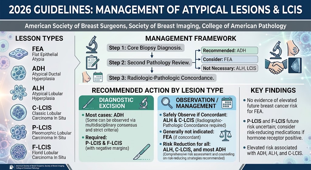
– Systematic risk assessment and counseling: Patients diagnosed with ADH, ALH, or C‑LCIS should receive comprehensive breast cancer risk assessment and counseling about risk-reducing strategies.
– Selective excision: Diagnostic excision is recommended for most ADH, P‑LCIS, and F‑LCIS identified on core biopsy; ALH and C‑LCIS may often be observed if imaging-pathology concordant. FEA alone typically does not require excision if concordant.
– Margin policy: P‑LCIS and F‑LCIS require negative margins at excision; ADH does not require a margin for excision (excision is diagnostic rather than oncologic).
– Pathology review: Mandatory second pathology review is advised for ADH; second review should be considered for FEA; not routine for ALH or LCIS when diagnosis is straightforward.
– Chemoprevention: Risk-reducing medications (eg, SERMs, aromatase inhibitors where appropriate) should be discussed for patients with ADH, ALH, or C‑LCIS. For P‑LCIS and F‑LCIS, chemoprevention is recommended when lesions are hormone receptor positive.
Updated Recommendations and Key Changes from Prior Practice
What’s new or clarified
– Clearer triage by lesion subtype: Prior guidelines and practices varied widely; the 2026 guideline provides lesion‑specific pathways (FEA, ADH, ALH, C‑LCIS, P‑LCIS, F‑LCIS) linked to imaging concordance and core‑biopsy features.
– Stronger emphasis on multidisciplinary decision-making: Radiology-pathology-surgery consensus is highlighted for selected ADH cases that may be observed rather than excised.
– Formalized role for second pathology review: ADH now carries a recommendation for confirmatory pathology review to reduce interobserver variability.
– Margin guidance introduced for LCIS variants: The requirement for negative margins with P‑LCIS and F‑LCIS is emphasized—an explicit surgical goal not previously standardized.
Evidence driving the updates
– Meta-analyses and pooled institutional series documenting variable but meaningful upgrade rates for lesions such as ADH and pleomorphic LCIS when sampled by core biopsy.
– Long-term cohort data demonstrating that ADH, ALH, and C‑LCIS confer an increased future breast cancer risk (e.g., Hartmann et al., NEJM 2005).
– Improved understanding of radiologic-pathologic concordance as a reliable filter to identify low‑risk lesions that can be observed safely.
Topic-by-Topic Recommendations
Note on levels of evidence and consensus: The 2026 guideline combines available evidence with expert consensus where data are limited. Recommendations are phrased with the guideline’s strength (evidence-informed consensus) rather than rigid numeric grades.
1) Pathology confirmation
– ADH: Recommend mandatory second pathology review or expert breast pathologist confirmation due to known interobserver variability.
– FEA: Consider second review when diagnosis is uncertain or when imaging-pathology concordance is unclear.
– ALH and LCIS: Second opinion not routinely required when classic features are present and the report is unambiguous.
2) Radiologic-pathologic concordance
– Concordance assessment is required for all core-biopsy diagnoses of atypia or LCIS. If the imaging abnormality is fully explained by the pathology finding, the lesion may be considered concordant.
– Discordant lesions should prompt diagnostic excision.
(Reference framework: ACR BI-RADS concepts applied at multidisciplinary conferences.)
3) Excision vs observation by lesion type
– FEA: If radiologic-pathologic concordant, diagnostic excision generally not indicated. Excise if discordant or if additional high‑risk features exist.
– ADH: Diagnostic excision recommended for most ADH identified on core biopsy. Selected ADH cases that meet strict criteria—very small focus, complete removal by vacuum‑assisted biopsy, imaging concordance, multidisciplinary consensus—may be observed with close imaging follow-up.
– ALH: May be observed if core biopsy diagnosis is concordant with imaging and there are no high‑risk imaging features. Excision recommended if discordant or if there are coexisting suspicious findings.
– C‑LCIS: Observational management is acceptable when radiologic-pathologic concordance is confirmed. Excision indicated for discordant results or when imaging shows mass or calcifications not explained by C‑LCIS.
– P‑LCIS and F‑LCIS: Diagnostic excision is recommended for all cases detected on core biopsy. At excision, negative margins should be achieved because these variants carry higher upgrade rates and more aggressive potential.
4) Margin policy
– ADH: No specific margin requirement; the aim of excision is diagnostic and to assess upgrade risk.
– P‑LCIS and F‑LCIS: Negative surgical margins recommended. If margins are positive or close, re-excision should be considered given higher upgrade and local recurrence risk.
5) Chemoprevention and risk reduction
– ADH, ALH, C‑LCIS: Patients should be counseled about chemoprevention (eg, tamoxifen, raloxifene, or aromatase inhibitors for postmenopausal patients) in accordance with established risk-reduction guidance (see USPSTF 2019). Shared decision-making is essential, considering comorbidity, menopausal status, and patient preferences.
– P‑LCIS and F‑LCIS: If lesions are hormone receptor positive, chemoprevention should be discussed and offered as appropriate.
– All patients with these lesions should be offered formal risk assessment (eg, Tyrer‑Cuzick or other validated models) and consideration for enhanced surveillance (annual mammography +/- tomosynthesis and, for selected women, MRI surveillance).
6) Surveillance
– Enhanced imaging surveillance is recommended for patients with ADH, ALH, or C‑LCIS given increased long‑term risk. MRI is individualized based on overall risk model estimates.
– For those observed after concordant ADH or ALH, short-interval imaging (eg, diagnostic mammography/ultrasound at 6 months) may be used before returning to standard annual screening if stable.
7) Special populations
– Young patients, pregnant or breastfeeding women, and patients with strong family history/genetic predisposition should be managed in multidisciplinary settings with individualized plans. Genetic counseling/testing should be considered when family history or other features suggest hereditary risk.
Expert Commentary and Insights
Panel perspective
– Multidisciplinary emphasis: The guideline committee underscored that management decisions should be made in multidisciplinary conferences whenever possible—melding imaging, pathologic, and surgical input reduces both unnecessary excisions and missed cancers.
– Balancing harm and benefit: Experts stressed that overuse of excisional surgery for low-risk lesions (eg, concordant FEA) can cause morbidity without clear benefit, whereas timely excision of P‑LCIS and F‑LCIS can prevent missed invasive cancers.
– Interobserver pathology variability: The recommendation for mandatory second reads of ADH reflects longstanding concerns about diagnostic reproducibility; expert breast pathologists improve diagnostic accuracy.
Areas of controversy and research needs
– ADH observation criteria: Although selected ADH may be observed, the exact thresholds (size, number of foci, completeness of removal) require prospective validation.
– Absolute risk estimates for P‑LCIS and F‑LCIS: Data remain limited; longitudinal studies are needed to better quantify subsequent invasive cancer risk.
– Optimal surveillance intervals: More data are needed to define the best imaging cadence for observed lesions.
Practical Implications for Clinical Practice
How the guideline changes day-to-day care
– Standardize preoperative workflows: Incorporate routine radiologic-pathologic concordance checks and a rapid pathway for second pathology review for ADH.
– Reduce unnecessary procedures: Expect fewer excisions for concordant FEA and some ALH/C‑LCIS cases, decreasing patient morbidity and resource use.
– Improve counseling and prevention: Routine risk assessment and a proactive discussion about chemoprevention will become standard for patients with ADH, ALH, and C‑LCIS.
– Surgical planning: When P‑LCIS or F‑LCIS is identified, plan for excision with the goal of negative margins and coordinate early multidisciplinary review.
Patient example
Maria Thompson, 54, underwent screening mammography with tomosynthesis showing clustered microcalcifications. Core biopsy returned focal ADH that the radiologist and pathologist agreed explained the imaging. Under previous practice she might have had automatic excision. Under the 2026 guideline, Maria’s case is discussed in a multidisciplinary conference; because the lesion is small, removed completely by vacuum‑assisted biopsy, and both imaging and pathology are concordant, the team offers observation with short-interval imaging and formal risk counseling including discussion of tamoxifen. Maria opts for surveillance and begins annual mammography with 6‑month diagnostic follow-up—avoiding surgery while accepting proactive monitoring.
References
– Nakhlis F, Bedrosian I, King TA, Heller SL, Allison KH, Boolbol S, Pass HA, Johnson NM, Boughey JC, Yao K. American Society of Breast Surgeons, Society of Breast Imaging, and College of American Pathology 2026 Guidelines for the Management of Proliferative Lesions With Atypia and Lobular Carcinoma In Situ. JAMA Surg. 2026 Aug 19. PMID: 42616513.
– Hartmann LC, Sellers TA, Frost MH, et al. Benign breast disease and the risk of breast cancer. N Engl J Med. 2005;353(3):229-237.
– US Preventive Services Task Force. Medication Use to Reduce Risk of Primary Breast Cancer in Women: Recommendation Statement. JAMA. 2019.
– American College of Radiology. ACR BI-RADS Atlas (5th ed.). Reston, VA: American College of Radiology; 2013.
– College of American Pathologists. Protocol for the Examination of Specimens From Patients With Invasive Breast Carcinoma and Ductal Carcinoma In Situ (2023 revisions). CAP; 2023.
Note: Readers should consult the full 2026 multisociety guideline for detailed algorithms, exclusion criteria for observation, and sample radiologic‑pathologic concordance scenarios. The guideline’s multidisciplinary approach is intended to improve consistency in management and to individualize care based on lesion subtype, imaging features, and patient preferences.