Highlights
- Quantitative CT imaging enables refined COPD phenotyping by assessing mucus plug burden and emphysema severity concurrently.
- The combined high mucus plug and high emphysema subtype is associated with the highest risk of acute exacerbations.
- Mucus plug burden independently predicts exacerbations especially in patients with low emphysema, underscoring distinct pathophysiological contributions.
- These imaging biomarkers facilitate personalized risk stratification and have potential utility in guiding targeted preventive interventions.
Background
Chronic obstructive pulmonary disease (COPD) is a heterogeneous lung disorder characterized primarily by airflow limitation and accompanied by varying degrees of emphysematous destruction and airway inflammation. Acute exacerbations of COPD (AECOPD) significantly impact morbidity, mortality, and healthcare utilization. Traditional clinical assessments and spirometry fail to sufficiently capture the biological and clinical heterogeneity underlying COPD, limiting prognostic accuracy and personalized management. Emerging quantitative computed tomography (CT) metrics offer promise in delineating disease phenotypes by objectively measuring structural changes such as emphysema extent and airway abnormalities, including mucus plugging. Understanding how these imaging biomarkers synergistically inform exacerbation risk could improve prognostic precision and tailor preventive therapies.
Key Content
Quantitative CT Metrics in COPD Phenotyping
Quantitative chest CT has evolved as a powerful noninvasive tool providing detailed assessment of lung parenchymal destruction and airway morphology. Emphysema extent is commonly quantified by the percentage of low attenuation areas below -950 Hounsfield units (%LAA-950), which correlates with histopathological emphysema severity and clinical outcomes. Concurrently, airway abnormalities including mucus plugs, luminal narrowing, and wall thickening can be quantified with emerging scoring methods. Mucus plugs reflect airway obstruction from secretions and contribute to ventilation-perfusion mismatch and exacerbation susceptibility.
Study Design and Patient Stratification
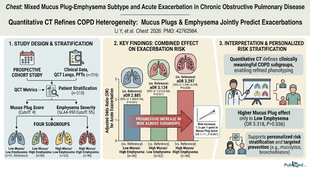
The referenced prospective cohort study by Li et al. (2026) enrolled 215 COPD patients who underwent comprehensive clinical evaluation, pulmonary function testing, and chest CT imaging. Patients were stratified by mucus plug score (cutoff 4) and emphysema severity (%LAA-950 cutoff 5%) into four subgroups: low mucus/low emphysema, low mucus/high emphysema, high mucus/low emphysema, and high mucus/high emphysema. This stratification allowed assessment of combined and individual contributions of mucus plugs and emphysema to acute exacerbation risk.
Association With Acute Exacerbations
Multivariate logistic regression adjusting for confounders revealed progressive increases in exacerbation risk across subgroups compared to low mucus/low emphysema: low mucus/high emphysema (adjusted OR 2.885), high mucus/low emphysema (OR 3.134), and high mucus/high emphysema (OR 3.297), all statistically significant. Notably, each 1-point increment in mucus plug score conferred a 1.1-fold increase in odds of exacerbation. Stratified analysis demonstrated that elevated mucus burden (≥4) predicts exacerbations significantly in patients with low emphysema but less so in those with high emphysema, suggesting differential mechanistic pathways.
Integration With Existing Literature
Previous studies have established emphysema and airway wall thickening individually associate with exacerbation risk; however, mucus plugging as a discrete imaging biomarker has been understudied. This study integrates these features, highlighting that mucus plugs contribute independently and augment risk beyond emphysema extent alone. The findings align with evidence implicating mucus hypersecretion and impaired clearance in COPD pathogenesis and exacerbation triggers. Also, they suggest potential phenotype-driven interventions such as mucolytics or airway clearance techniques might serve patients with predominant mucus plug phenotype.
Methodological Advances
The use of standardized quantitative CT metrics for mucus plug scoring and emphysema measurement marks an advancement over qualitative visual assessments, reducing observer bias and allowing reproducible phenotyping. The cutoffs used enable clinically applicable subgroup delineation. The prospective cohort design and multivariate analysis reinforce the validity of findings. Nevertheless, further validation in larger, diverse cohorts and longitudinal studies will strengthen generalizability.
Expert Commentary
This landmark study underscores the utility of refined CT-based phenotyping in COPD, moving beyond traditional spirometric metrics toward imaging biomarkers that reflect underlying pathobiology. The identification of a mixed mucus plug-emphysema subtype offers a nuanced perspective on COPD heterogeneity with genuine prognostic implications. Clinically, these findings advocate for integrating quantitative CT analysis into routine evaluation for enhanced risk stratification.
However, practical implementation requires access to standardized CT protocols, image analysis expertise, and consideration of radiation exposure. Importantly, the mucus plug metric’s association with exacerbation only within the low emphysema subgroup suggests that tailored preventive strategies might differ between phenotypes—patients with high mucus plugging may benefit from therapies aiming at mucus clearance and anti-inflammatory approaches targeting airway obstruction, whereas emphysema-predominant patients might require other modalities.
Biologically, mucus plugs exacerbate ventilation heterogeneity and foster microenvironmental conditions favorable for bacterial colonization and exacerbation initiation. These mechanistic insights highlight mucus plugs not merely as markers but as therapeutic targets. Complementary investigations integrating molecular biomarkers, sputum profiling, and longitudinal exacerbation phenotyping could further delineate causal pathways.
Conclusion
Quantitative CT-derived assessment of mucus plugs combined with emphysema severity robustly identifies COPD subgroups with differential exacerbation risk. The mixed mucus plug-emphysema phenotype exhibits the highest risk, and mucus plug burden independently predicts acute exacerbations particularly in patients with lesser emphysema. These findings pave the way for personalized risk stratification and targeted prevention strategies in COPD, emphasizing the clinical relevance of refined quantitative imaging biomarkers. Future research should focus on validation, longitudinal assessment, and interventional trials tailored to phenotypic subgroups to translate imaging phenotypes into improved patient outcomes.
References
- Li Y, Wang Y, Zhang M, Wang G, Cui J, Zhang Z, Wang X, Yang X, Guo Y. Mixed Mucus Plug-Emphysema Subtype and Acute Exacerbation in Chronic Obstructive Pulmonary Disease. Chest. 2026 Sep 19. PMID: 42762984.
- Matsuoka S, et al. Quantitative CT assessment of airway disease and emphysema in COPD. Proc Am Thorac Soc. 2008;5(9):906-911. PMID: 19017931.
- Han MK, et al. Chronic obstructive pulmonary disease phenotypes. Am J Respir Crit Care Med. 2010;182(5):598-604. PMID: 20651237.
- Vestbo J, Hurd SS, Agustí AG, et al. Global Strategy for the diagnosis, management, and prevention of chronic obstructive pulmonary disease GOLD executive summary. Am J Respir Crit Care Med. 2013;187(4):347-365. PMID: 22878278.
- McDonough JE, et al. Small-airway obstruction and emphysema in chronic obstructive pulmonary disease. N Engl J Med. 2011;365(17):1567-1575. PMID: 22047508.