Highlights
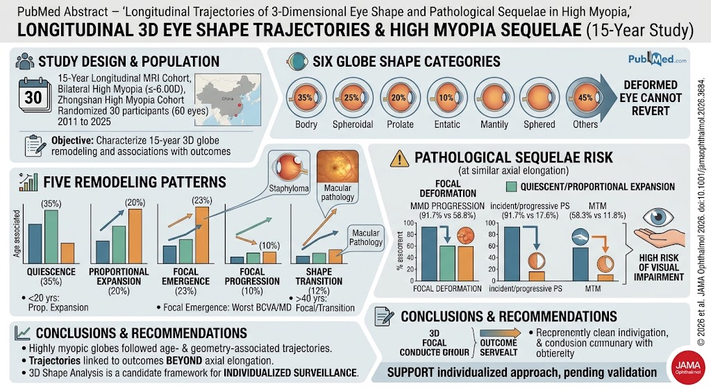
- Highly myopic eyes remodel in 3D along distinct longitudinal trajectories defined by age and baseline globe morphology.
- Five remodeling patterns—quiescence, proportional expansion, focal emergence, focal progression, and shape transition—associate variably with visual deterioration and pathological progression.
- 3D shape deformation, especially focal deformation patterns, correlates strongly with increased risk of myopic macular degeneration (MMD), posterior staphyloma (PS), and myopic traction maculopathy (MTM), independently of axial elongation.
- Baseline 3D globe shape subtypes provide valuable prognostic stratification and support the use of MRI-based eye shape analysis as a candidate tool for personalized surveillance and intervention planning.
Background
High myopia, typically defined as spherical equivalent refractive error ≤ -6.00 diopters, affects a growing global population and is a major cause of irreversible vision loss due to progressive pathological changes in the posterior segment of the eye. Traditional clinical risk stratification relies heavily on axial length measurement; however, recent evidence highlights the complex three-dimensional remodeling of the globe contributing to complications such as myopic macular degeneration (MMD), posterior staphyloma (PS), and myopic traction maculopathy (MTM). Despite recognition of 3D eye shape as an important factor, longitudinal trajectories and their relationship with clinical outcomes have remained incompletely characterized.
Key Content
Chronological and Cohort Evidence on 3D Eye Shape Remodeling Trajectories
Chen et al. (2026) reported a landmark 15-year longitudinal substudy within the Zhongshan High Myopia Cohort involving 30 participants (60 eyes) with bilateral high myopia, assessed by high-resolution 3D MRI in 2011 and followed till 2025. Using an innovative classification scheme encompassing six globe shapes (spheroidal, ellipsoidal, conical, nasally distorted, temporally distorted, and barrel-shaped), five remodeling patterns were defined: quiescence (no shape change), proportional expansion, focal emergence, focal progression, and shape transition.
Pattern distribution was age-dependent: younger individuals (<20 years) predominantly exhibited proportional expansion, indicative of global symmetric enlargement, while older subjects (≥40 years) were more prone to focal progression or shape transition, involving localized morphological deformities. Importantly, no eye with initial deformation regressed to a spheroidal shape, underscoring the progressive nature of deformation. This study also linked remodeling patterns to clinical outcomes: eyes with focal emergence had the greatest declines in best-corrected visual acuity (0.26 logMAR) and visual field mean deviation (-3.50 dB). Those undergoing shape transition exhibited significant visual field pattern standard deviation increases (1.95 dB).
Association of Eye Shape with Pathological Sequelae Beyond Axial Length
At similar axial elongation rates, eyes developing focal deformations had significantly higher risks of MMD progression (91.7% vs. 58.8%; P=0.02), incident or progressive PS (91.7% vs. 17.6%; P<0.001), and MTM (58.3% vs. 11.8%; P=0.003) compared to those with non-deformed or proportionally expanded eyes. These findings highlight that 3D shape change adds prognostic value beyond axial length alone.
Corresponding studies reinforce the prognostic significance of 3D eye shape. A prospective cohort analysis by Li et al. (2026) involving 95 participants showed that nasally distorted and conical eye shapes had the fastest axial length elongation and the highest risks of macular choroidal thinning and MMD progression. Deformed eyes overall exhibited a sevenfold increase in macular choroidal thinning risk, with visual field defects also more prevalent in deformed morphologies.
Further, a 4-year longitudinal study (Wang et al., 2025) demonstrated that eye shape deformity independently predicted myopic maculopathy progression (OR 4.35) similarly to axial length ≥28 mm (OR 12.75). Incorporating eye shape with demographic and biometric data improved predictive accuracy (AUC 0.862).
Morphological Spectrum and Clinical Implications
Cross-sectional analyses confirm a spectrum of eye shape deformations linked to pathology severity. Spheroidal (non-deformed) eyes predominate in younger cohorts and associate with milder MMD and better visual acuity, while barrel-shaped, temporally or nasally distorted eyes, and eyes with PS correlate with more advanced myopic degeneration and visual impairment (Chen et al., 2017). This underlines the role of focal globe contour abnormalities in exacerbating biomechanical stress and chorioretinal atrophy.
Expert Commentary
The accumulated evidence establishes 3D eye shape remodeling as a dynamic and multifaceted process influenced by age and inherent globe geometry. These shape changes are not merely epiphenomena of axial elongation but confer independent risk for sight-threatening complications and visual function decline.
Integration of MRI-based 3D shape classification into routine clinical care remains exploratory but offers promising potential for personalized risk stratification. The stability of deformation patterns over 15 years, and their predictive value for MMD, PS, and MTM progression, advocate for their incorporation into surveillance algorithms. However, practical challenges include accessibility, cost, and standardization of 3D MRI acquisition and analysis.
Biologically, focal deformation may represent localized mechanical weaknesses or remodeling in the scleral shell, precipitating staphyloma formation and secondary retinal pathology. This biomechanical perspective aligns with observed phenotype-specific risks and suggests potential targets for future interventional therapies aimed at biomechanical reinforcement.
Guidelines have yet to integrate 3D eye shape assessment formally, but emerging longitudinal data from the Zhongshan cohorts provide compelling impetus to develop MRI-based prognostic tools, possibly combined with axial length, choroidal thickness, and functional assessments to optimize management.
Conclusion
Over the past decade and recently extended to 15 years, strong evidence from prospective and longitudinal cohorts confirms that highly myopic eyes undergo distinct 3D shape remodeling trajectories that correlate with key pathological sequelae. These remodeling patterns carry prognostic significance beyond traditional axial length measurements and visual acuity alone.
Future directions should focus on multicenter validation of 3D shape classification systems, development of non-MRI surrogate imaging modalities, and clinical trials testing interventions tailored to specific shape phenotypes. Ultimately, 3D eye shape analysis promises to enhance individualized clinical surveillance and improve outcomes for patients with high myopia.
References
- Chen S, Li Y, Xuan M, et al. Longitudinal Trajectories of 3-Dimensional Eye Shape and Pathological Sequelae in High Myopia. JAMA Ophthalmol. 2026 Sep 3:e263684. PMID: 42690636.
- Li Y, Chen S, Xuan M, et al. Pathologic Myopia Globe Shape and Long-Term Prognosis. JAMA Ophthalmol. 2026 Jul 1;144(7):608-617. PMID: 42207540.
- Wang W, et al. Eye Shape Deformity Predicts Myopic Maculopathy Progression Among Highly Myopic Individuals: A 4-Year Longitudinal Study. Retina. 2025 Jan;45(1):52-60. PMID: 39699857.
- Chen S, et al. Three-Dimensional Eye Shape, Myopic Maculopathy, and Visual Acuity: The Zhongshan Ophthalmic Center-Brien Holden Vision Institute High Myopia Cohort Study. Ophthalmology. 2017 May;124(5):679-687. PMID: 28237427.