Highlights
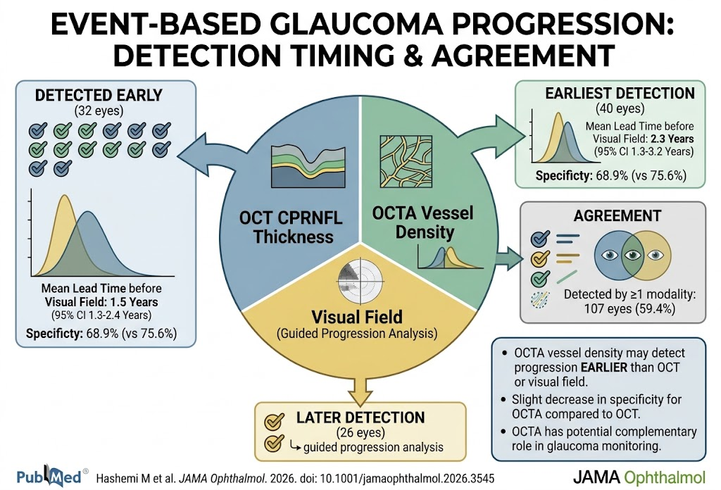
- Optical coherence tomography angiography (OCTA) vessel density detects glaucoma progression approximately 2.3 years earlier than visual field (VF) loss and 0.2 years earlier than OCT circumpapillary retinal nerve fiber layer (CPRNFL) thinning.
- OCTA demonstrates slightly lower specificity compared to OCT for event-based progression detection, suggesting a complementary rather than a replacement role.
- Baseline microvascular abnormalities such as microvasculature dropout (MvD) and choroidal microvascular dropout (CMvD) are associated with faster progression, underscoring vascular contributions to glaucoma pathophysiology.
- Advanced analytical methods including machine learning and deep learning applied to longitudinal OCTA imaging show promise in improving progression prediction accuracy.
Background
Glaucoma remains a leading cause of irreversible blindness worldwide, characterized by progressive optic neuropathy and corresponding visual field loss. Early detection of disease progression is critical to preserve vision and tailor treatment intensity. Conventional clinical monitoring combines structural measurement of retinal nerve fiber layer (RNFL) thinning using optical coherence tomography (OCT) with functional assessment through standard automated perimetry (visual field testing). However, these methods often detect progression only after significant damage has occurred. Optical coherence tomography angiography (OCTA), a non-invasive imaging technique that quantifies retinal and optic nerve head microvasculature, has emerged as a novel tool potentially capable of earlier detection of glaucomatous damage by revealing microvascular changes preceding structural loss or functional decline.
Key Content
Event-Based Progression Detection: Comparative Timing and Agreement
The landmark retrospective study by Hashemi et al. (2026) evaluated 180 eyes from 116 patients followed for a mean of 5.1 years with concurrent OCT, OCTA, and VF testing. Event-based progression was defined for OCT and OCTA as changes surpassing test-retest variability at two consecutive visits, while VF progression employed guided progression analysis. Among eyes showing progression (59.4%), OCTA vessel density changes detected progression earliest in 37.4%, compared to 29.9% by OCT CPRNFL thickness and 24.3% by VF. The lead time of OCTA preceding VF progression was 2.3 years (95% CI 1.3–3.2), and OCT preceded VF by 1.5 years (95% CI 0.6–2.4). Specificity was higher for OCT (75.6%) than OCTA (68.9%). These findings position OCTA vessel density as a valuable early biomarker of glaucomatous progression, though with modest compromise in specificity.
Microvascular Dropout and Its Prognostic Value
Baseline microvasculature dropout (MvD) detected on OCTA predicts faster capillary density loss and subsequent VF deterioration in preperimetric glaucoma (PPG) eyes, as demonstrated by Shin et al. (2026). This vascular deficiency correlates with a higher risk of conversion from structural appearance to functional loss. Similarly, choroidal microvascular dropout (CMvD) progression relates to more rapid VF decline, with differences observed between primary open-angle glaucoma (POAG) and pseudoexfoliation glaucoma (PXG). Such vascular features may serve as prognostic markers to stratify risk and guide monitoring frequency.
Reproducibility and Variability Across Modalities
Measurement variability is critical when interpreting progression. Wu et al. (2026) reported that VF and OCTA metrics exhibit severity-dependent variability, with VF mean deviation (MD) tolerance limits doubling in moderate-to-advanced glaucoma and OCTA peripapillary perfusion density showing variable coefficients of variation between 5% and 9%. Conversely, OCT structural measures like peripapillary RNFL and macular ganglion cell-inner plexiform layer (mGCIPL) thickness maintained stable variability (~2%) across glaucoma severities. This evidence underscores the need for modality-specific and disease-stage adjusted thresholds for progression detection.
Structural and Vascular Correlates in Advanced Disease
In advanced glaucoma, common structural measures may approach a floor effect limiting their monitoring utility. Yu et al. (2025) demonstrated that macular parafoveal superficial capillary plexus vessel density (SCP-VD) correlates strongly with central 10-2 VF loss and ganglion cell complex thickness, suggesting OCTA vascular parameters complement structural metrics and can serve as surrogate markers particularly when VF testing reliability is limited.
Impact of Myopia and Glaucoma Subtypes
Highly myopic glaucomatous eyes show distinct progression patterns. Mori et al. (2025) found optic disc vessel density (ODVD) reduction strongly associated with VF progression in high myopia, whereas RNFL thinning was less predictive. Meanwhile, Yen et al. (2026) highlighted that normal-tension glaucoma (NTG) patients below 40 years with high myopia and reduced macular vessel density exhibited faster progression. These findings emphasize that vascular metrics are particularly informative in myopic and NTG subpopulations.
Advanced Analytical Techniques
Artificial intelligence approaches have been applied to longitudinal OCTA data to improve progression detection. Wen et al. (2024) developed a deep learning model using longitudinal en-face macular OCTA images which achieved an area under the curve (AUC) of 0.81, outperforming logistic regression based on vessel density alone. Furthermore, machine learning classifiers incorporating structural and vascular parameters, such as deep vascular plexus density and choriocapillaris dropout, have been used to predict progression rates with high sensitivity and specificity (Chen et al., 2025).
Therapeutic Interventions Targeting Vascular Dysregulation
Adjunctive therapies aiming to improve ocular blood flow, such as systemic dronabinol administration, have shown increased optic nerve head blood flow and superficial vessel density on OCTA in primary open-angle glaucoma (POAG) patients without affecting intraocular pressure (IOP) (Muller et al., 2026). These interventions highlight the translational implications of vascular biomarkers in both monitoring and therapeutic paradigms.
Expert Commentary
Despite OCT and VF remaining the diagnostic mainstays for glaucoma monitoring, evidence increasingly supports incorporating OCTA vascular metrics into clinical practice to detect progression earlier. The trade-off between earlier detection and slightly reduced specificity necessitates careful clinical judgment to avoid overtreatment. Vascular parameters may be especially valuable when structural OCT approaches floor effects or when VF testing is unreliable.
Moreover, vascular dysfunction likely contributes to glaucoma pathogenesis and progression, as supported by microvasculature dropout associations and differential retinal vasoreactivity in NTG versus high-tension glaucoma. These insights reinforce the rationale for vascular-targeted therapies.
Nevertheless, limitations include measurement variability, the need for standardized progression criteria for OCTA, and cost-effectiveness considerations of routine OCTA in diverse clinical settings. Future research should focus on prospective, multicenter validations, refinement of normative databases, and integration with artificial intelligence for personalized monitoring.
Conclusion
Emerging evidence from longitudinal cohorts and sophisticated analytical models affirms that OCTA vessel density metrics offer earlier detection of glaucoma progression compared to OCT structural and visual field testing. These vascular biomarkers enrich the glaucoma monitoring armamentarium, informing individualized risk stratification and potentially guiding novel therapeutic approaches targeting ocular perfusion. Meticulous clinical integration of OCTA requires balancing sensitivity, specificity, and economic factors. Continued research will delineate optimal strategies for leveraging multimodal imaging to improve visual outcomes in glaucoma patients.
References
- Hashemi M, Nishida T, Moghimi S, et al. Event-Based Progression Detection Among OCT, OCT Angiography, and Visual Field in Glaucoma. JAMA Ophthalmol. 2026; e263545. PMID:42690635
- Shin JW, Sung KR, Park SB, et al. Progression in Preperimetric Glaucoma Eyes With and Without Microvasculature Dropout. Am J Ophthalmol. 2026;289:224-230. PMID:42214583
- Wu Z, Wen J, Rao HL, et al. Reproducibility of VF, OCT, and OCTA in Glaucoma: A Prospective Multi-Visit Study. Transl Vis Sci Technol. 2026;15(7):30. PMID:42524829
- Yu H, Su H, Zhao J, et al. Optic Disc Microvasculature Reduction and Visual Field Progression in Advanced Primary Open-Angle Glaucoma. Am J Ophthalmol. 2026;287:163-177. PMID:41912062
- Mori S, Ikuno Y, Yoshikawa M, et al. Longitudinal Measurement of Optic Disc Vessel Density to Detect Glaucoma Progression in High Myopia. Ophthalmology. 2025;132(12):1357-1371. PMID:40769299
- Wen JC, Zhao B, Schuman JS, et al. Detection of glaucoma progression on longitudinal series of en-face macular optical coherence tomography angiography images with a deep learning model. Br J Ophthalmol. 2024;108(12):1688-1693. PMID:39117359
- Chen Y, Li J, Shao Y, et al. Predictive modeling of glaucomatous optic neuropathy progression rate using OCT and OCTA parameters. Vestn Oftalmol. 2025;141(2):22-29. PMID:40353537
- Muller PL, Dettmann J, Siedlecki J, et al. Single oral administration of dronabinol increases ocular blood flow in patients with glaucoma: A randomized controlled trial. Acta Ophthalmol. 2026;104(2):225-232. PMID:40772417