Highlight
1. Oral epithelial dysplasia (OED) is a precursor lesion with variable malignant transformation risk, currently assessed by subjective histological grading.
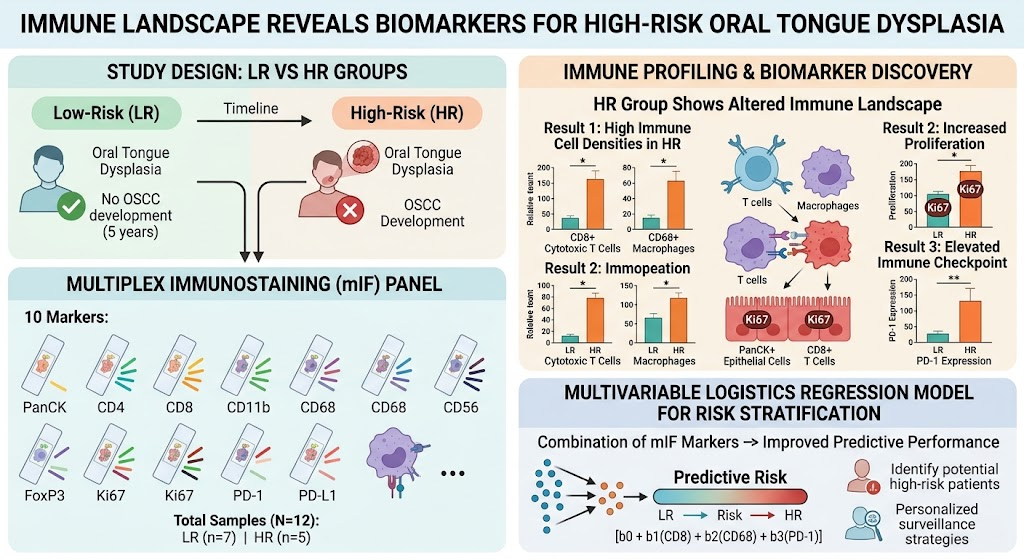
2. Multiplex immunostaining differentiates low-risk (LR) and high-risk (HR) oral tongue dysplasia based on immune landscape.
3. The HR group shows increased CD8+ cytotoxic T cells, CD68+ macrophages, and immunoinhibitory PD-1 expression.
4. Proliferation marker Ki67 is elevated in epithelial and CD8+ cells in HR cases, and a multimarker logistic model enhances predictive accuracy.
Study Background
Oral epithelial dysplasia (OED) represents histopathologic alterations in oral mucosa epithelium indicating increased cancer risk, particularly for progression to oral squamous cell carcinoma (OSCC). The oral tongue is a common site for OED with high malignant transformation potential. Clinical management relies primarily on histological gradings, such as binary (low vs. high grade) or tertiary classification systems (mild, moderate, severe dysplasia). However, these gradings are subject to substantial interobserver variability and limited predictive value. Identification of robust biomarkers to reliably segregate high-risk from low-risk OED cases is an unmet clinical need to guide surveillance and therapeutic interventions.
Study Design
This study included 12 archival oral tongue dysplasia samples from 12 patients, grouped into low-risk (LR, n=7) who did not develop OSCC within five years and high-risk (HR, n=5) who progressed to OSCC. Multiplex immunostaining employed a panel of 10 antibodies targeting markers of epithelial cells (PanCK), T cell subsets (CD4, CD8), myeloid cells (CD11b, CD68), natural killer cells (CD56), regulatory T cells (FoxP3), proliferation (Ki67), and immune checkpoints (PD-1, PD-L1). Computational analysis through Uniform Manifold Approximation and Projection (UMAP) assessed differences in immune populations and functional states between LR and HR groups. Logistic regression modeling evaluated individual marker predictive value and developed a multimarker risk stratification model.
Key Findings
Distinct immune landscapes segregated LR from HR oral tongue dysplasia cases. The HR group demonstrated a significantly higher infiltration of CD8+ cytotoxic T lymphocytes and CD68+ macrophages as compared to the LR group. Ki67, a marker of cellular proliferation, was elevated in both PanCK+ epithelial cells and CD8+ T cells in the HR group, suggesting increased proliferative activity not just in dysplastic epithelial cells but also in cytotoxic immune cells.
Immune checkpoint analysis revealed significantly increased PD-1 expression in the HR group, indicating an immunosuppressive microenvironment facilitating malignant progression. PD-L1 expression differences were less pronounced. The presence of PD-1+ immune cells may point towards exhaustion or dysfunction of antitumor lymphocytes during tumorigenesis.
Logistic regression identified individual markers—CD8+ cell density, CD68+ macrophage count, and PD-1 expression—as predictors of progression risk. A multimarker logistic model combining these variables outperformed any single marker in discriminating HR from LR cases, with improved sensitivity and specificity consistent with clinical risk stratification needs.
Expert Commentary
This study offers novel insights into the immunological alterations underlying malignant transformation risk in oral tongue dysplasia. The paradoxical increase in proliferating CD8+ T cells in high-risk lesions may reflect an initially robust cytotoxic immune response that becomes functionally impaired through PD-1 mediated exhaustion. Macrophages, particularly CD68+ populations, may contribute to a pro-tumor microenvironment through immunomodulatory and trophic roles. These findings align with emerging concepts that immune contexture critically influences premalignant lesion fate.
While promising, limitations include the small sample size and retrospective design. Future validation in larger cohorts and longitudinal studies is needed to confirm the clinical utility of these biomarkers. Integration with histological grading could address existing subjectivity and refine patient surveillance paradigms. Additionally, functional studies may elucidate the mechanisms by which these immune subsets influence transformation, potentially revealing therapeutic targets for immunoprevention.
Conclusion
This multiplex immunostaining study identifies a distinct immune signature comprising proliferative CD8+ T cells, CD68+ macrophages, and elevated PD-1 expression that associates with high-risk oral tongue dysplasia. These immune biomarkers hold promise for objectively stratifying patients at increased malignant transformation risk beyond conventional histology. Implementing such biomarkers in clinical practice could optimize individualized follow-up and early intervention strategies, ultimately improving oral cancer outcomes.
Funding and ClinicalTrials.gov
The original article did not specify funding or registered clinical trial information.
References
Sandell A, Giacomini M, Risteli M, Kilpinen S, Salo T, Al-Samadi A. Immune Landscape Reveals Biomarkers for High-Risk Oral Tongue Dysplasia. J Dent Res. 2026;105(11):1462-1470. PMID: 41834374.
Additional references on OED and immunology may include:
1. Speight PM, et al. Oral epithelial dysplasia classification and grading: Development and use in clinical practice. J Pathol. 2018;244(5):580-591.
2. Schütz E, et al. Immune microenvironment in oral squamous cell carcinoma: A review. Front Oncol. 2020;10:570924.
3. Gandhi S, et al. The role of immune checkpoint inhibitors in head and neck cancers: Current evidence and future perspectives. Oral Oncol. 2017;73:99-105.