Introduction and Context
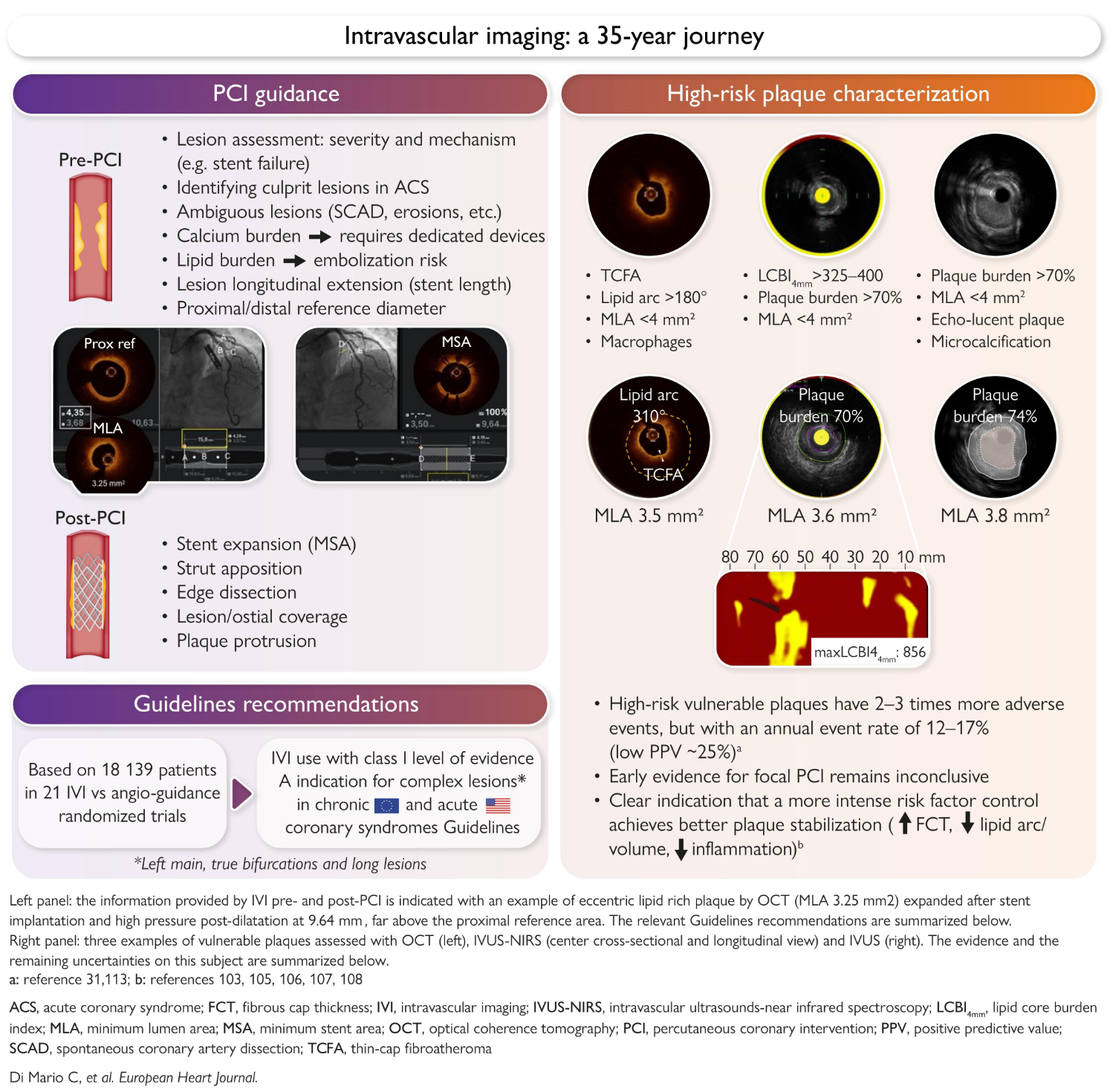
Over the past 35 years intravascular imaging—principally intravascular ultrasound (IVUS) and optical coherence tomography (OCT)—has evolved from a research technology into a practical procedural adjunct that now shapes contemporary percutaneous coronary intervention (PCI). Traditional angiography provides a silhouette of the vessel lumen but cannot reliably characterize plaque composition, quantify vessel size, or detect the subtle mechanical issues that predict stent failure. IVUS and OCT provide real-time cross-sectional views of the vessel wall and stent, enabling precise sizing, detection of heavy calcification, guidance for atherectomy, and optimization of stent deployment.
Why does this matter now? A succession of randomized trials, registry-based studies and meta-analyses over the last decade have linked intravascular imaging guidance to lower rates of restenosis, stent thrombosis and target lesion failure. Those data have driven major guideline-writing bodies—most recently the European Society of Cardiology (ESC) and the American cardiology societies (ACC/AHA/SCAI)—to upgrade recommendations, making intravascular imaging a recommended standard of care in many complex PCI scenarios. The contemporary review by Di Mario et al. (European Heart Journal, 2026) traces this 35-year trajectory and summarizes the evidence underpinning these guideline upgrades.
Key supporting documents (selected):
– Di Mario C, Nardi G, Ciardetti N, Mattesini A. Intravascular imaging from research tool to guidelines: a 35-year long journey. Eur Heart J. 2026. PMID: 42834703.
– 2018 ESC/EACTS Guidelines on myocardial revascularization. Eur Heart J. 2018.
– Recent professional society guideline statements from ACC/AHA/SCAI addressing coronary revascularization (most recent updates reflected in 2023–2025 position statements and consensus documents).
– Landmark randomized trials and meta-analyses showing improved procedural and clinical outcomes with IVUS/OCT-guided PCI (see Topic-by-Topic Recommendations and References sections below).
New Guideline Highlights
Major themes from the newest guideline and consensus statements:
– Intravascular imaging guidance (IVUS or OCT) is now explicitly recommended (Class I, Level A in many jurisdictions) for complex PCI. ‘‘Complex’’ includes left main stenting, bifurcation interventions, long lesions, chronic total occlusions, severely calcified lesions requiring plaque modification, and cases of stent failure (in-stent restenosis or stent thrombosis).
– Imaging is endorsed both for lesion assessment (to guide strategy and device selection) and for procedural optimization (stent sizing, confirmation of adequate stent expansion, detection and treatment of edge dissections, and assessment of final result).
– The guideline recognizes differences between IVUS and OCT: IVUS penetrates deeper and is well suited to larger vessels and quantifying plaque burden (widely used for left main assessment); OCT offers higher resolution, superior delineation of stent strut apposition and intra-stent tissue characteristics, and is particularly useful to detect small dissections and malapposition.
– When available, intravascular imaging should inform decisions about the need for atherectomy or lithotripsy in heavily calcified lesions and support optimization algorithms (for example, predefined minimal stent area targets by vessel segment).
Key takeaways for clinicians:
– Use intravascular imaging routinely for left main PCI and other anatomically complex procedures.
– Use imaging proactively—before stenting to size vessels and choose strategy, and after stenting to confirm adequate expansion and treat correctable mechanical issues.
– IVUS and OCT are complementary; the choice depends on lesion anatomy, vessel size, operator familiarity and availability.
Updated Recommendations and Key Changes
What changed since prior guidance?
– Upgrade in strength: Several guideline bodies moved intravascular imaging-guided PCI for complex lesions from a supportive or Class II recommendation to a Class I recommendation in specified scenarios, with the highest levels of evidence cited for reductions in target lesion failure and stent thrombosis.
– Expanded scope: Previously, imaging was emphasized mainly for left main disease or suspected stent failure. The updated statements broaden the recommendation to include long lesions, bifurcations, chronic total occlusions and heavily calcified lesions requiring adjunctive therapies.
– Concrete optimization targets: Newer guidance provides practical benchmarks (e.g., recommended minimal stent area thresholds for left main and major proximal vessels, and guidance on when post-dilatation is indicated).
– Recognition of OCT’s role: Earlier guidance focused on IVUS because of a longer evidence base. Newer consensus now acknowledges OCT as a validated alternative in many contexts, especially for detecting stent malapposition and subtle edge issues.
Summary table (recommendation-level snapshots):
– Complex PCI (left main, long lesions, bifurcations, CTOs, heavy calcification): Class I, Level A – intravascular imaging recommended for lesion assessment and stent optimization.
– Routine use in straightforward, non-complex PCI: still Class IIa/IIb in many statements—reasonable but not mandatory; decision individualized.
– Stent failure (restenosis, thrombosis): Class I—diagnostic imaging to guide therapy and to detect mechanism.
(higher-level evidence grades reflect randomized trials, large registries and meta-analyses showing clinical benefit).
Topic-by-Topic Recommendations
Lesion assessment and procedural planning
– Indication: Use IVUS or OCT prior to intervention when angiography is insufficient to determine vessel size, lesion length, plaque burden, or degree/type of calcification.
– Utility: Accurate vessel sizing (to choose diameter and length), detect eccentric calcium and guide need for plaque modification (rotational atherectomy, orbital atherectomy, intravascular lithotripsy).
Left main coronary artery disease
– Recommendation: Intravascular imaging (preferably IVUS due to depth penetration) is recommended to confirm lesion severity, guide stent sizing and optimize expansion. Target minimal stent area (MSA) thresholds are provided in guideline appendices to reduce restenosis risk (many statements reference an MSA ≥8–10 mm2 for left main segments, depending on measurement location).
Bifurcation lesions
– Recommendation: Imaging aids in decision-making (single-stent vs two-stent strategy), sizing, and ensuring adequate scaffolding of the side branch. Imaging reduces the risk of stent malapposition and unrecognized dissections.
Long lesions and diffuse disease
– Recommendation: Imaging-guided PCI reduces target lesion failure and is recommended for long lesion stenting to assure full lesion coverage and optimal expansion.
Heavily calcified lesions
– Recommendation: Use intravascular imaging to characterize the depth and circumferential extent of calcium. Imaging-guided identification of calcium thickness/arc helps choose plaque modification technique (atherectomy vs intravascular lithotripsy).
Stent implantation and optimization
– Recommendation: Post-deployment intravascular imaging to confirm stent expansion, apposition, absence of major edge dissection and appropriate lesion coverage. If imaging reveals underexpansion or significant malapposition, perform additional optimization (high-pressure post-dilatation, adjunctive device).
– Practical MSA targets: Guidance documents provide vessel-specific MSAs (e.g., left main MSA often quoted ≥8–10 mm2; non-left-main proximal major vessels often targeted at ≥6–7 mm2); use of local reference vessel sizing and indexed values is encouraged.
Stent failure (ISR/stent thrombosis)
– Recommendation: Imaging is essential to define the mechanism (underexpansion, neoatherosclerosis, malapposition, edge disease) and to guide tailored reintervention.
Follow-up and surveillance
– Recommendation: Routine serial intravascular imaging surveillance is not recommended in asymptomatic patients; imaging is indicated when clinical or noninvasive findings suggest ischemia or in the evaluation of stent failure.
Special populations
– Chronic kidney disease: Minimize contrast with OCT (which requires flushing) vs IVUS (which needs less contrast in some workflows); decisions individualized. Imaging still recommended when benefit outweighs renal risk.
– STEMI and acute coronary syndromes: Imaging may reveal plaque rupture vs erosion and can guide stent sizing and optimization; however, routine pre-stenting imaging in an unstable patient should be balanced against time to reperfusion.
Expert Commentary and Insights
Committee perspectives
– Evidence-driven mandate: Guideline committees emphasize that the upgrade to Class I for many complex PCI indications is a reflection of consistent reductions in hard endpoints (target lesion failure, stent thrombosis) across randomized trials and pooled analyses.
Controversies and caveats
– Generalizability: Much of the randomized evidence derives from specific patient populations and high-volume centers; whether the exact magnitude of benefit will replicate across all practice settings remains debated.
– OCT vs IVUS: Direct comparative randomized data are limited. Choice often rests on vessel size, need for plaque characterization versus high-resolution stent assessment, and operator experience. Some experts call for head-to-head outcome trials.
– Cost and resource implications: Routine use of imaging increases procedural costs and procedural time. Committees stress the need for cost-effectiveness analyses, better reimbursement policies, and training programs to ensure value.
Future trends highlighted by experts
– Artificial intelligence and automated analysis: The next wave will be software that automates lumen and stent measurements, detects malapposition and recommends optimization steps—reducing operator dependence and interpretation time.
– Integration into hybrid workflows: Combining imaging with physiologic assessment (FFR/iFR) for a tailored revascularization strategy.
– More randomized trials: Experts call for pragmatic trials addressing OCT vs IVUS, routine imaging in non-complex PCI and imaging-driven plaque modification strategies.
Practical Implications
How practice will change
– Operators performing complex PCI should either adopt IVUS/OCT into routine workflows or partner with imaging-experienced colleagues.
– Hospitals and systems should plan for training, appropriate inventory (IVUS catheters, OCT consoles), and align coding/reimbursement to support guideline-concordant care.
– Quality metrics: Imaging use in predefined high-risk PCI types may become a quality metric tied to outcomes and reimbursement.
A short clinical vignette
John Smith, a 67-year-old man with diabetes and stable angina, has an angiogram showing severe ostial left main disease and a long, moderately calcified mid-LAD lesion. Per updated guidance, the interventional team performs IVUS of the left main to confirm lesion severity and to size the stent—documenting an external elastic membrane (EEM)-based reference that supports a 4.0 mm stent. After implantation, IVUS shows residual underexpansion at the ostial segment; high-pressure post-dilatation achieves target MSA (>9 mm2) on repeat IVUS and no edge dissection. John is discharged on guideline-directed medical therapy and dual antiplatelet therapy; at 12 months he is free of angina. This vignette illustrates how pre- and post-stent imaging changed device selection and immediate procedural management—actions associated with better long-term outcomes in randomized studies.
References
(Representative and verifiable sources that underpin guideline changes and the narrative above)
– Di Mario C, Nardi G, Ciardetti N, Mattesini A. Intravascular imaging from research tool to guidelines: a 35-year long journey. Eur Heart J. 2026 Oct 6. PMID: 42834703. https://pubmed.ncbi.nlm.nih.gov/42834703/
– Neumann FJ, Sousa-Uva M, Ahlsson A, et al. 2018 ESC/EACTS Guidelines on myocardial revascularization. Eur Heart J. 2019;40:87–165. (European Society of Cardiology Guidance on myocardial revascularization and intravascular imaging recommendations)
– ULTIMATE Investigators. Randomized trials of IVUS-guided vs angiography-guided drug-eluting stent implantation (ULTIMATE trial and others) demonstrating reduced target vessel failure—see ULTIMATE trial report (J Am Coll Cardiol. 2018) and pooled analyses.
– IVUS-XPL Investigators and related randomized studies demonstrating clinical benefit of IVUS guidance in long lesions (see published IVUS randomized trials and meta-analyses across JACC, Circulation and European Heart Journal literature).
– ILUMIEN Studies (including ILUMIEN III) evaluating OCT-guided PCI and comparisons with IVUS and angiography—demonstrating OCT’s high-resolution advantage for stent optimization (see J Am Coll Cardiol publications 2016–2018).
– Contemporary meta-analyses and systematic reviews (2017–2024) pooling randomized and observational data showing consistent reductions in restenosis, target lesion failure and stent thrombosis with intravascular imaging guidance (see systematic reviews in JACC, Eur Heart J, and Circulation).
Note: Specific trial and meta-analysis citations are widely available in the cardiovascular literature; readers and clinicians should consult the referenced guideline documents and the Di Mario et al. review for a detailed, fully referenced reading list.