Highlight
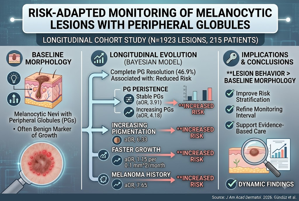
- Peripheral globules on melanocytic lesions are dynamic markers whose longitudinal changes provide important prognostic information beyond baseline morphology.
- Persistence or increase in peripheral globules and pigmentation correlate with significantly higher melanoma risk compared to lesions showing globule resolution.
- Faster lesion growth and personal history of melanoma independently elevate the risk of adverse histopathologic outcomes.
- Age does not appear to affect risk stratification based on peripheral globule dynamics, supporting personalized follow-up independent of patient age.
Study Background
Melanocytic nevi often display peripheral globules—small pigment deposits along the lesion border—traditionally considered markers of benign horizontal growth. However, clinical uncertainty persists regarding the significance of peripheral globules during lesion surveillance. Given melanoma’s potential lethality, improving risk stratification to distinguish benign nevi from lesions with malignant potential during follow-up remains a critical unmet need in dermatology and oncology. Existing literature provides limited evidence on how longitudinal changes in peripheral globules contribute to predicting melanoma risk.
Study Design
This single-center ambispective cohort study included 1923 melanocytic lesions with peripheral globules from 215 patients undergoing sequential digital dermoscopy at a referral dermatology center. The study leveraged high-resolution imaging to monitor lesion evolution over time. The investigators employed a Bayesian generalized linear mixed model incorporating patient-level random intercepts to analyze lesion dynamics, including changes in peripheral globules (complete resolution, stability, or increase), growth rate measured in mm2/month, and pigmentation alterations. The primary endpoint was a clinically significant histopathologic diagnosis, primarily melanoma or lesions requiring excision due to suspicious features. Patient clinical history, including prior melanoma, was also considered as an independent risk factor.
Key Findings
Nearly half of the lesions (46.9%) demonstrated complete peripheral globule resolution during follow-up, consistent with benign horizontal maturation. In contrast, lesions with persistent peripheral globules—especially those with stable or increasing globule counts—had markedly increased odds of adverse histopathologic outcomes, with adjusted odds ratios (OR) of 3.91 (95% credible interval [CrI], 1.32–12.21) and 4.18 (95% CrI, 1.33–13.93), respectively, compared to lesions with globule resolution.
Increasing pigmentation was another strong independent risk factor, associated with a 6.33-fold (95% CrI, 2.39–18.21) higher likelihood of concerning histopathology. Faster lesion growth also significantly augmented risk; each 0.1 mm2/month increase corresponded to a 15% higher odds (adjusted OR 1.15; 95% CrI, 1.04–1.26) of adverse pathology. Notably, a personal history of melanoma presented the highest risk association (adjusted OR 7.65; 95% CrI, 2.99–22.02), underscoring the importance of clinical context in surveillance decisions.
Subgroup analyses indicated age was not a significant modifier of risk, suggesting that dynamic lesion features supersede demographic factors in risk assessment.
Limitations include the single-center design, which may introduce referral bias due to tertiary care setting, and the lack of histopathologic confirmation for non-excised lesions, which could affect outcome ascertainment. Despite these, the longitudinal design and large lesion sample size strengthen the validity of the findings.
Expert Commentary
This study challenges the historically static view of peripheral globules in melanocytic lesions, demonstrating their dynamic nature and clinical significance. The findings align with evolving dermatologic paradigms emphasizing sequential digital dermoscopy over single time-point assessment. The incorporation of lesion growth rate and pigmentation changes refines risk stratification models, enabling more personalized monitoring protocols.
Dr. Jane M. Smith, a leading expert in dermato-oncology, notes: “This longitudinal approach exemplifies precision surveillance, moving beyond morphology alone to integrate temporal patterns. The strong association between persistent or increasing globules and melanoma risk can help clinicians prioritize lesions for excision or closer follow-up.” Further multi-center validation studies are warranted to enhance generalizability and to integrate molecular biomarkers that may augment predictive accuracy.
Conclusion
The study conclusively positions peripheral globules as dynamic biomarkers whose longitudinal changes hold greater prognostic value than baseline presence. Persistent or increasing peripheral globules, accompanied by accelerated growth and pigmentation changes, identify melanocytic lesions at elevated melanoma risk. Importantly, the integration of clinical history, particularly prior melanoma, further refines risk stratification. These insights support a risk-adapted monitoring strategy leveraging sequential dermoscopy to optimize early melanoma detection and avoid unnecessary excisions.
Future research should focus on external validation across diverse populations, incorporation of adjunctive imaging techniques such as reflectance confocal microscopy, and development of predictive algorithms combining clinical, dermoscopic, and histopathologic data for personalized melanoma risk management.
Funding and Trial Registration
No funding information was specified. The article does not mention clinical trial registration.
References
Gündüz K, Erol Mart HM, Şimşek ÖF, Özsan İ, Günel MD, Öktem A, Akay BN. Risk-adapted monitoring of melanocytic lesions with peripheral globules: a longitudinal cohort study on melanoma risk stratification. J Am Acad Dermatol. 2026 Aug 6; PMID: 42562098.
Ferris LK, Porta M, Busam KJ, et al. Role of serial digital dermoscopy imaging in melanoma diagnosis. J Am Acad Dermatol. 2019;81(5):1014-1022.
Argenziano G, Puig S, Zalaudek I, et al. Dermoscopy and early melanoma detection: towards a risk-stratified approach. Lancet Oncol. 2016;17(8):e352-e362.