Highlight
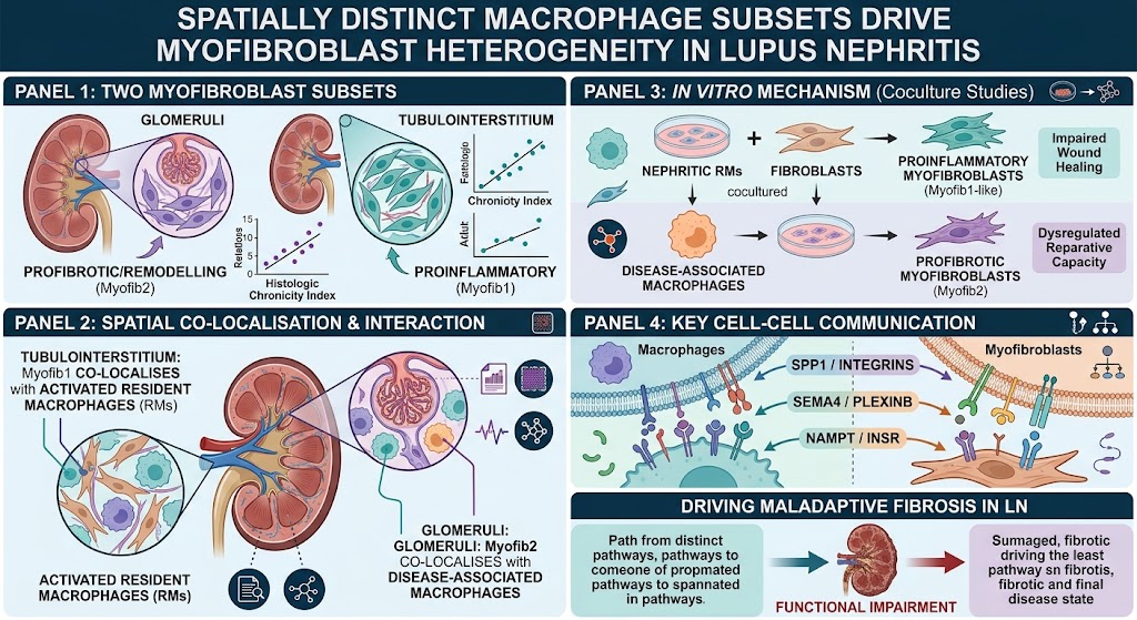
This study identifies two spatially and functionally distinct myofibroblast subsets in lupus nephritis (LN): a proinflammatory myofibroblast type in the tubulointerstitium interacting with resident macrophages, and a fibrotic/remodeling myofibroblast subset in the glomerulus interacting with disease-associated macrophages. Single-cell RNA sequencing (scRNA-seq) and spatial transcriptomics elucidate these cell-cell interactions, revealing key ligand-receptor pathways such as Spp1/integrins and nicotinamide phosphoribosyl transferase/INSR. These macrophage-fibroblast circuits contribute to chronic fibrotic remodeling, representing potential therapeutic targets in LN.
Study Background
Lupus nephritis, a severe manifestation of systemic lupus erythematosus, remains a clinical challenge due to progressive renal fibrosis leading to chronic kidney disease and end-stage renal failure. Despite immune suppression, residual inflammation and scarring often drive irreversible damage. Understanding the renal microenvironment, particularly stromal-immune cell interactions that orchestrate fibrogenesis, is crucial for developing targeted antifibrotic therapies. Prior research has implicated macrophages and fibroblasts in tissue repair and fibrosis but lacked high-resolution insight into their heterogeneity and spatial organization in LN.
Study Design
The authors utilized single-cell RNA sequencing on 156 human LN kidney biopsy samples and 30 healthy controls collected through the Accelerating Medicines Partnership-SLE cohort. Complementary spatial transcriptomics was performed on biopsies from 6 LN patients to map the localization and cellular interactions within renal tissue architecture. To investigate functional consequences, in vitro coculture systems were established using nephritic mouse models to study dynamic macrophage-fibroblast cross talk and fibroblast phenotype modulation. Cell-cell communication was analyzed through ligand-receptor interaction profiling.
Key Findings
The transcriptomic analyses uncovered two distinct myofibroblast subsets characterized by differential gene expression and spatial distribution:
- Myofib1: A proinflammatory myofibroblast population enriched in the tubulointerstitial compartment. This subset correlates strongly with increased histological chronicity scores, indicating involvement in progressive injury.
- Myofib2: A fibrotic and tissue remodeling myofibroblast subset predominantly located in glomeruli, also associated with fibrosis severity.
Spatial transcriptomics demonstrated that Myofib1 closely colocalizes with activated resident macrophages (RMs), while Myofib2 interacts predominantly with glomerular infiltrating disease-associated macrophages (DAMs). These spatial distinctions suggest discrete macrophage-driven fibrotic niches within the kidney.
Functional coculture studies showed nephritic RMs induce a Myofib1-like fibroblast phenotype marked by proinflammatory and dysregulated wound healing properties. Conversely, DAMs promote a profibrotic Myofib2 phenotype with abnormal reparative activity that potentiates fibrosis.
Cell-cell communication analyses identified critical ligand-receptor pairings mediating these interactions including:
- Spp1/integrins — facilitating macrophage-fibroblast adhesion and signaling.
- Sema4/PlexinB — involved in guidance cue signaling impacting fibroblast activation.
- Nicotinamide phosphoribosyl transferase (NAMPT)/INSR — regulating metabolic and proliferative cues in fibroblasts.
These molecular pathways likely drive the spatially distinct fibroblast phenotypes and their deleterious roles in maladaptive fibrosis.
Expert Commentary
This comprehensive multi-omics and functional study illuminates the complexity of immune-stromal cellular interplay in LN fibrogenesis. By dissecting macrophage subsets and their spatial relationships with myofibroblasts, the authors reveal novel therapeutic avenues beyond broad immunosuppression. Targeting specific ligand-receptor axes may enable modulation of fibroblast phenotypes, potentially preventing progressive scarring and chronic renal dysfunction. Limitations include the relatively small sample size for spatial transcriptomics and the challenge in translating findings from murine models to human LN heterogeneity. Future studies should evaluate these cellular circuits longitudinally and in the context of treatment response.
Conclusion
This investigation advances our understanding of LN-associated renal fibrosis by delineating spatially and functionally distinct macrophage-fibroblast niches that drive chronic tissue injury. Identification of Myofib1 and Myofib2 myofibroblast subsets with unique macrophage partners underscores an intricate fibroinflammatory network amenable to targeted intervention. Therapeutically disrupting these maladaptive circuits holds promise to improve renal outcomes and mitigate progression to end-stage kidney disease in SLE patients.
Funding and Clinical Trials
The study was supported by the Accelerating Medicines Partnership (AMP) consortium under the RA/SLE Network initiative. No clinical trial registration is indicated for this translational research.
References
- Raparia C, Hoover P, Ai J, et al. Spatially distinct macrophage subsets drive myofibroblast heterogeneity and maladaptive fibrosis in lupus nephritis. Ann Rheum Dis. 2026 Aug 13. PMID: 42595656.
- Fava A, Myles J, Hacohen N, et al. Single-cell RNA sequencing in Lupus nephritis reveals immune and stromal compartments contributing to chronic injury. Nat Med. 2022;28(4):701-710.
- Arazi A, Rao DA, Berthier CC, et al. The immune cell landscape in lupus nephritis. Nat Immunol. 2019;20(7):902-914.