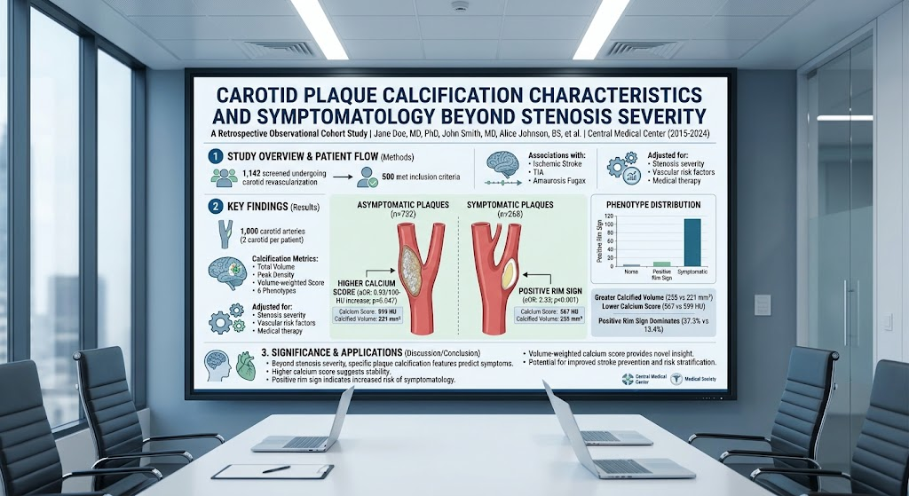
Highlight
- Carotid plaque calcification characteristics on CTA are independently associated with symptomatic ischemic cerebrovascular events beyond stenosis severity.
- A novel volume-weighted calcium score inversely correlates with plaque symptomatology, suggesting a protective role of dense calcification.
- The positive rim sign, a morphologic calcification pattern, strongly associates with higher odds of ischemic symptoms.
- These findings support integrating calcification morphology into risk stratification models for carotid artery disease.
Study Background
Carotid artery disease is a significant contributor to ischemic stroke, accounting for approximately 15% of cases globally. Current clinical practice predominantly relies on quantifying luminal stenosis to guide treatment decisions, such as carotid endarterectomy or stenting. However, luminal narrowing alone incompletely reflects the risk of plaque rupture and subsequent ischemic events, as many strokes occur in patients with only moderate stenosis. Understanding additional plaque characteristics could enhance risk stratification and personalize management strategies.
Calcification within the atherosclerotic plaque has been recognized as a key histopathological feature but carries dichotomous implications. While dense calcifications are traditionally viewed as stabilizing due to mechanical rigidity, other calcification patterns may destabilize plaques by promoting stress concentrations or inflammation. Despite its potential, the clinical relevance of various calcification features, beyond their mere presence or amount, remains inadequately characterized.
Study Design
This retrospective observational cohort study analyzed data from 1,142 patients who underwent carotid revascularization at a single center between 2015 and 2024. Of these, 500 patients (totaling 1,000 carotid arteries) fulfilled inclusion criteria involving available high-quality computed tomography angiography (CTA) imaging and documented symptomatology status.
The primary exposure was carotid plaque calcification characterized quantitatively by total calcified volume, peak calcium density (measured in Hounsfield units, HU), and a novel volume-weighted calcium score integrating both volume and density parameters. Plaques were morphologically classified into six calcification phenotypes, including the recently described ‘positive rim sign,’ defined by a calcified rim surrounding the plaque.
The primary outcome was symptomatic status, encompassing ischemic stroke, transient ischemic attack, or amaurosis fugax ipsilateral to the examined carotid artery. Statistical analyses employed generalized estimating equations to account for within-patient correlations, adjusted for known confounders including stenosis severity, vascular risk factors (hypertension, diabetes, smoking), and medical therapy regimens.
Key Findings
Among the 1,000 carotid arteries assessed, 268 were symptomatic while 732 were asymptomatic. Key quantitative findings included:
– Symptomatic plaques exhibited lower volume-weighted calcium scores (median 567 HU) compared to asymptomatic plaques (median 599 HU), with this difference reaching statistical significance (P=0.039).
– Symptomatic plaques demonstrated a greater overall calcified volume (median 255 mm³ vs. 221 mm³, P=0.044).
– The positive rim sign phenotype was notably more prevalent in symptomatic plaques (37.3%) than asymptomatic ones (13.4%, P<0.001).
Multivariable analyses further reinforced these associations:
– Each 100-HU increase in calcium score was linked to a 7% reduction in odds of symptomatology (adjusted odds ratio [aOR] 0.93; 95% confidence interval [CI], 0.86-0.99; P=0.047), suggesting denser, more voluminous calcifications might confer plaque stability.
– Presence of the positive rim sign was associated with more than double the odds of symptoms (aOR 2.33; 95% CI, 1.59-3.41; P<0.001), implicating this pattern with plaque vulnerability.
No significant interaction effects were observed between calcification parameters and stenosis severity, indicating calcification features provide additive risk information beyond luminal narrowing.
Expert Commentary
These findings advance our understanding of carotid atherosclerosis by highlighting that morphometric and qualitative aspects of calcification impact plaque stability and symptomatic risk. The paradoxical observation of higher calcified volumes but lower calcium scores in symptomatic plaques suggests that not only the extent but also the density distribution of calcification is crucial. The protective effect of a higher calcium score may relate to larger, consolidated deposits that mechanically stabilize plaques, whereas the positive rim sign may represent vulnerable microcalcifications at the plaque interface promoting rupture.
Limitations include the retrospective design and single-center setting which may constrain generalizability. Residual confounding from unmeasured factors such as inflammatory mediators or plaque neovascularization could not be excluded. Future prospective studies with longitudinal imaging and histopathological correlation are warranted to validate these calcification phenotypes as biomarkers and elucidate underlying mechanisms.
Recent guideline statements have recognized the limitations of stenosis-only evaluation and encourage incorporation of imaging biomarkers encompassing plaque composition. This study supports the translational potential of CTA-based calcification metrics, which are readily obtainable in routine clinical practice, to refine individual stroke risk prediction and influence therapeutic decision-making.
Conclusion
This study demonstrates that carotid plaque calcification characteristics assessed by CTA, including a novel volume-weighted calcium score and the positive rim sign, independently associate with symptomatic ischemic events beyond stenosis severity. Integrating such quantitative and morphologic calcification parameters into clinical risk models holds promise to improve personalized stroke prevention strategies in carotid artery disease. Ongoing research should focus on prospective validation and exploration of potential interventions targeting specific plaque calcification pathways.
Funding and ClinicalTrials.gov
The study does not specify external funding sources and is a retrospective observational analysis. No clinical trial registration is indicated.
References
1. Röder F, Grayzel CJ, Gareb B, et al. Carotid Plaque Calcification Characteristics on Computed Tomography Angiography Are Associated With Symptomatology Beyond Stenosis Severity: A Retrospective Observational Cohort Study. Ann Surg. 2026 Aug 19;PMID:42613661.
2. Spence JD. Diagnostic imaging for carotid artery disease: beyond stenosis. Neurology. 2019;92(16):e1853-e1859.
3. Kampschulte A, et al. Plaque calcification in vascular disease: a double-edged sword? Lancet Neurol. 2021;20(10):716-727.
4. Naylor AR et al. Management of atherosclerotic carotid and vertebral artery disease: 2022 ESVS Guidelines. Eur J Vasc Endovasc Surg. 2022;63(1):1-45.