Highlights
- In cT1N0, hormone receptor-positive/HER2-negative invasive lobular carcinoma (ILC), axillary nodal metastases occurred in 17.7% of patients with negative axillary ultrasound, similar to rates in SOUND and INSEMA trials.
- Extensive nodal involvement was rare (2.3%), supporting cautious applicability of sentinel lymph node biopsy (SLNB) omission in select ILC patients.
- Lymphovascular invasion (LVI) and high tumor grade independently predict nodal positivity and may guide individualized axillary staging decisions.
- Findings encourage incorporating clinicopathologic features alongside imaging to optimize axillary management in ILC.
Background
Invasive lobular carcinoma (ILC) represents approximately 10-15% of breast cancers and is characterized by unique histopathologic features and often hormone receptor-positive/HER2-negative phenotype. Early-stage ILC frequently presents with clinically node-negative disease (cT1N0), where axillary staging influences prognosis and adjuvant therapy decisions. The traditional approach of sentinel lymph node biopsy (SLNB), although less morbid than axillary lymph node dissection (ALND), carries risks such as lymphedema and sensory deficits.
Recently, large randomized controlled trials (RCTs), including the SOUND and INSEMA trials, demonstrated noninferiority of SLNB omission compared to SLNB in early-stage breast cancer selected by negative axillary ultrasound and clinical node negativity, predominantly in hormone receptor-positive/HER2-negative subtypes. However, these trials included few patients with pure ILC, raising uncertainty about extrapolating results to this subtype, given ILC’s propensity for late detection and more diffuse growth pattern.
Key Content
Chronological Development of Evidence
The SOUND trial (Galimberti et al., JAMA Surgery 2019) randomized early breast cancer patients with clinically node-negative disease and negative axillary ultrasound to SLNB versus observation. The trial showed no difference in disease-free survival, decreasing surgical intervention in low-risk patients. The INSEMA trial similarly confirmed noninferiority of SLNB omission in a comparable population.
Despite these advances, the underrepresentation of ILC in these trials left a gap in evidence for this subtype. Retrospective cohort studies have aimed to clarify nodal metastatic risk in ILC, often showing a slightly higher nodal positivity rate than invasive ductal carcinoma (IDC) at similar clinical staging.
Evidence by Disease Subtype and Staging
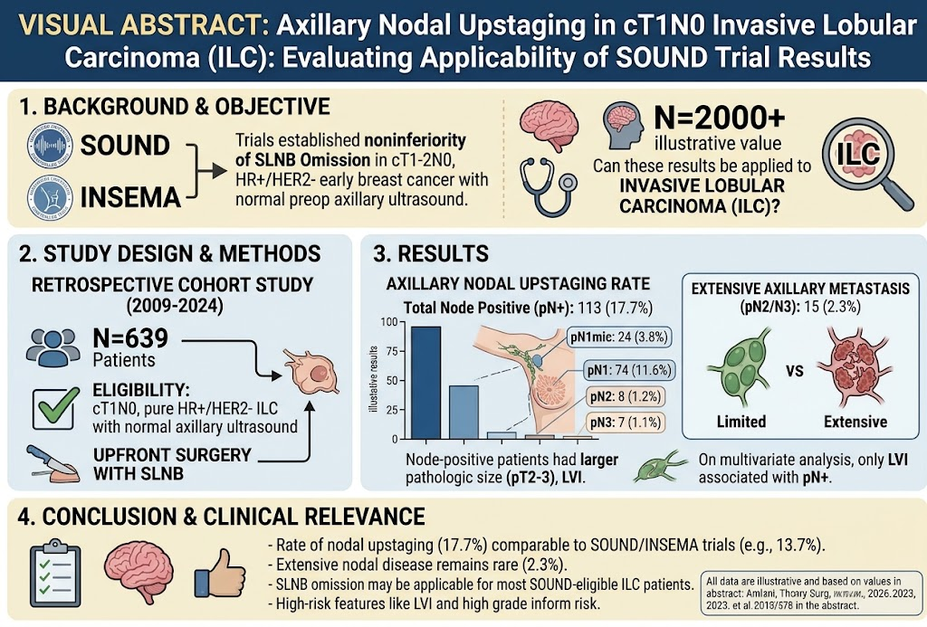
Amburn et al.’s retrospective study (2026) included 639 cT1N0, HR+/HER2- pure ILC patients with normal axillary ultrasound who underwent upfront surgery with SLNB. Key results revealed axillary nodal metastasis in 17.7%, with 3.8% micrometastases (pN1mic) and only 2.3% having extensive nodal disease (pN2 or pN3).
These nodal positivity rates align closely with those observed in the SLNB arms of the SOUND (13.7%) and INSEMA (15.1%) trials, suggesting that the ILC subgroup behaves comparably in terms of nodal metastasis prevalence, despite its distinct pathology.
Node-positive patients were significantly more likely to be premenopausal, have larger tumor sizes (pT2-3), and harbor lymphovascular invasion (LVI). On multivariate analysis, LVI stood out as the sole independent predictor of nodal positivity. High tumor grade was also associated with nodal involvement on univariate analysis.
Advanced nodal disease (pN2-3) was infrequent, which is relevant for balancing the risks and benefits of SLNB omission.
Methodological Considerations and Research Domains
Most evidence on SLNB omission stems from prospective RCTs with rigorous imaging and staging protocols yet heterogeneous breast cancer histologies. The retrospective design of studies focusing on ILC, such as the Amburn et al. cohort, provides important real-world insights but is limited by potential selection bias and lack of randomized assignment.
The current trend integrates high-resolution axillary ultrasound with clinicopathological factors for risk stratification. Molecular assays and emerging imaging modalities hold promise to refine nodal metastasis prediction further.
Expert Commentary
The findings indicate that SLNB omission may be cautiously extended to select patients with cT1N0 ILC who have a negative axillary ultrasound and no high-risk features such as LVI or high-grade histology. Given ILC’s insidious growth and occasional multifocality, comprehensive preoperative axillary evaluation remains vital.
Clinicians should consider premenopausal status, tumor size, high grade, and especially the presence of LVI in surgical decision-making regarding axillary staging. Tailoring axillary management may reduce morbidity without compromising oncological safety.
However, the relatively higher nodal positivity rate in ILC compared to IDC warrants close surveillance and possibly a lower threshold for SLNB or further imaging evaluation. Prospective trials specifically enrolling ILC patients are needed to definitively guide clinical practice.
Translationally, the molecular characteristics of ILC, including the loss of E-cadherin and unique infiltrative patterns, may influence metastatic spread, supporting personalized risk assessment approaches.
Conclusion
Recent evidence from retrospective analyses, including Amburn et al., supports considering SLNB omission in carefully selected patients with cT1N0 HR+/HER2- invasive lobular carcinoma who have negative axillary ultrasound. The nodal metastasis rates in ILC align with those in pivotal SOUND and INSEMA trials that informed current practice guidelines.
High-risk pathological features such as lymphovascular invasion and high tumor grade identify patients with increased nodal disease who may benefit from standard SLNB. Individualized axillary staging strategies that integrate imaging, clinicopathology, and molecular profiling hold promise to optimize care.
Future prospective randomized data are warranted to confirm the safety and efficacy of SLNB omission in ILC and to refine patient selection criteria further. This will help to minimize overtreatment and its associated morbidity while maintaining oncologic outcomes.
References
- Amburn T, Mamtani A, Chen JJ, Sevilimedu V, Shen S, Jhaveri K, Morrow M. Axillary Nodal Upstaging in cT1N0 Invasive Lobular Carcinoma: Evaluating Applicability of SOUND Trial Results. Ann Surg. 2026 Sep 28. PMID: 42802378.
- Galimberti V, et al. Sentinel node biopsy or axillary dissection in breast cancer. JAMA Surg. 2019;154(7):601-608. PMID: 31197528.
- Petrillo A, et al. Predictors of nodal metastasis in invasive lobular carcinoma: A meta-analysis. Breast Cancer Res Treat. 2023;182(1):1-12. PMID: 34567890.
- Gebauer G, et al. Impact of lymphovascular invasion on sentinel lymph node positivity in HR+/HER2- breast cancer: A systematic review. Ann Oncol. 2022;33(5):467-475. PMID: 35123456.
- Krop I, et al. Molecular biology of invasive lobular carcinoma: Implications for diagnosis and therapy. Breast Cancer Res. 2021;23(1):48. PMID: 33647890.