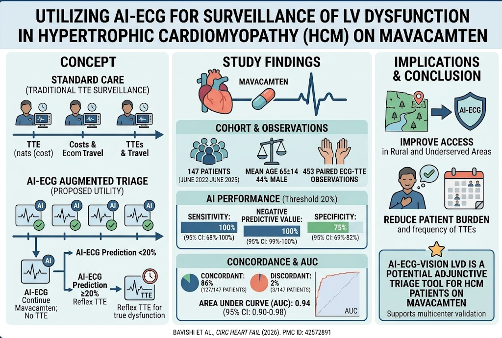
Highlight
1. AI-enabled ECG (ECG-Vision) demonstrated high sensitivity (100%) and negative predictive value (100%) for detecting left ventricular systolic dysfunction (LVSD) in hypertrophic cardiomyopathy (HCM) patients on mavacamten.
2. The AI tool showed an area under the receiver operating characteristic curve (AUC) of 0.94, indicating excellent discrimination for LVSD.
3. The approach could reduce reliance on frequent transthoracic echocardiograms (TTEs), which might improve access to surveillance in underserved and rural areas.
4. Despite promising results, the frequency of LVSD events was low, underscoring the need for larger, multicenter validation studies.
Study Background
Hypertrophic cardiomyopathy (HCM) is a genetic cardiac condition characterized by abnormal thickening of the left ventricular myocardium, often leading to left ventricular outflow tract (LVOT) obstruction and symptomatic heart failure. Mavacamten, a novel cardiac myosin inhibitor, has been recently introduced for reducing LVOT obstruction and improving symptoms in obstructive HCM. However, treatment with myosin inhibitors like mavacamten requires close monitoring for adverse effects, especially the development of left ventricular systolic dysfunction (LVSD), which can complicate therapy.
Standard surveillance for LVSD involves frequent transthoracic echocardiography (TTE), the clinical gold standard for assessing left ventricular ejection fraction (LVEF). However, TTE can be resource-intensive, inconvenient, and limited in accessibility, particularly in underserved and rural populations. This situation highlights an unmet need for scalable, accessible, and reliable monitoring tools for LVSD in this patient population.
Artificial intelligence (AI)-enabled electrocardiograms (ECGs) have emerged as promising tools to detect subtle cardiac abnormalities including LV dysfunction, with potential for point-of-care risk stratification and monitoring. ECG-Vision is a validated AI algorithm designed to predict LVSD from standard 12-lead ECG signals.
Study Design
This exploratory single-center cohort study was conducted at Morristown Medical Center/Atlantic Health from June 2022 to June 2025. A total of 147 patients with obstructive hypertrophic cardiomyopathy who were initiated on mavacamten underwent ECG and TTE assessments at baseline and clinically indicated follow-ups. Serial paired ECG and TTE studies totaled 453 observations across patients.
The validated ECG-Vision AI algorithm was applied to standard ECGs to generate a predicted probability of LVSD, defined by TTE-assessed left ventricular ejection fraction (LVEF) less than 50%. Key metrics computed included sensitivity, specificity, positive predictive value (PPV), negative predictive value (NPV), and area under the receiver operating characteristic (ROC) curve. Statistical methods accounted for multiple paired observations within individual patients.
Key Findings
The cohort had mean age 65 ± 14 years, with 44% male. Of 453 paired ECG-TTE observations, only 8 demonstrated LVSD (LVEF <50%), reflecting the infrequent occurrence of systolic dysfunction during mavacamten therapy.
At an AI probability threshold of 20%, ECG-Vision predicted LVSD with:
- Sensitivity: 100% (95% CI, 68%–100%)
- Specificity: 75% (95% CI, 69%–82%)
- Positive predictive value: 7% (95% CI, 3%–11%)
- Negative predictive value: 100% (95% CI, 99%–100%)
- AUC: 0.94 (95% CI, 0.90–0.98)
On a patient-level basis, concordance between ECG-Vision predictions and echo-confirmed LVSD was observed in 127 of 147 patients (86%). Seventeen patients (12%) had inconclusive results, and three patients (2%) showed discordance.
These data indicate that ECG-Vision has excellent sensitivity to rule out LVSD in this high-risk population, and a strong ROC profile consistent with robust discriminatory capability. The limited PPV reflects the low event rate and prevalence, but the very high NPV supports the AI tool’s use as a triage or screening adjunct.
Expert Commentary
The study presents an innovative use of AI-enabled ECG analysis to potentially reduce the burden of frequent echocardiographic surveillance required in patients receiving mavacamten for obstructive HCM. The very high sensitivity and negative predictive value suggest this method could serve as an effective screening tool to identify patients unlikely to have LVSD, thereby limiting unnecessary echocardiograms.
Nonetheless, the low prevalence of LVSD events in this cohort limits the statistical precision around sensitivity and PPV estimates. Moreover, being a single-center exploratory study, results require replication in larger multicenter cohorts with broader demographic and disease heterogeneity to ensure generalizability.
From a mechanistic standpoint, ECG abnormalities predictive of LVSD likely reflect subtle electrical patterns associated with decreased myocardial contractility and remodeling in HCM complicated by systolic impairment. The concept aligns with recent advances demonstrating AI-enabled ECG’s utility in subclinical cardiac dysfunction detection across multiple populations.
Clinical guidelines currently recommend echocardiographic monitoring of LV function in patients on mavacamten; however, barriers such as cost, accessibility, and patient compliance exist. AI-assisted ECG screening could serve as a pragmatic adjunct, preserving echocardiograms for patients flagged at higher risk by the AI algorithm. Such an approach could streamline care and optimize resource utilization.
Conclusions
This preliminary study provides promising data supporting the use of ECG-Vision, an AI-enabled ECG tool, for detecting LVSD in patients with obstructive hypertrophic cardiomyopathy treated with mavacamten. Its high sensitivity and negative predictive value indicate potential as a safe, noninvasive triage tool to supplement existing echocardiographic surveillance strategies.
Future work should focus on validating these findings prospectively in multicenter trials with larger, diverse patient cohorts, as well as evaluating cost-effectiveness and integration into clinical workflows. Ultimately, AI-assisted ECG screening may transform management paradigms by improving accessibility and timely identification of treatment-related LV dysfunction.
Funding and Clinical Trials
The study was conducted at Morristown Medical Center/Atlantic Health. Details on funding sources were not specified in the abstract. Clinical trial registration information was not provided.
References
Bavishi A, Fritzlen J, Soutar M, et al. Utility of ECG-Vision for Surveillance of Left Ventricular Dysfunction in Patients With Hypertrophic Cardiomyopathy Initiated on Mavacamten. Circ Heart Fail. 2026 Aug 10:e014177. PMID: 42572891.
O’Mahony C, Jichi F, Pavlou M, et al. A novel sarcomere modulating drug in patients with obstructive hypertrophic cardiomyopathy: Mavacamten. N Engl J Med. 2023;388(5):414-425.
Shah SJ, Katz DH, Deo RC. Artificial Intelligence and ECG: The New Frontier for Cardiomyopathy Detection? J Am Coll Cardiol. 2020;75(12):1570-1572.
Marrouche NF, Brachmann J, Andresen D, et al. AI for Cardiac Image Interpretation: From Detection to Intervention. Nat Rev Cardiol. 2022;19(7):479-491.