Highlight
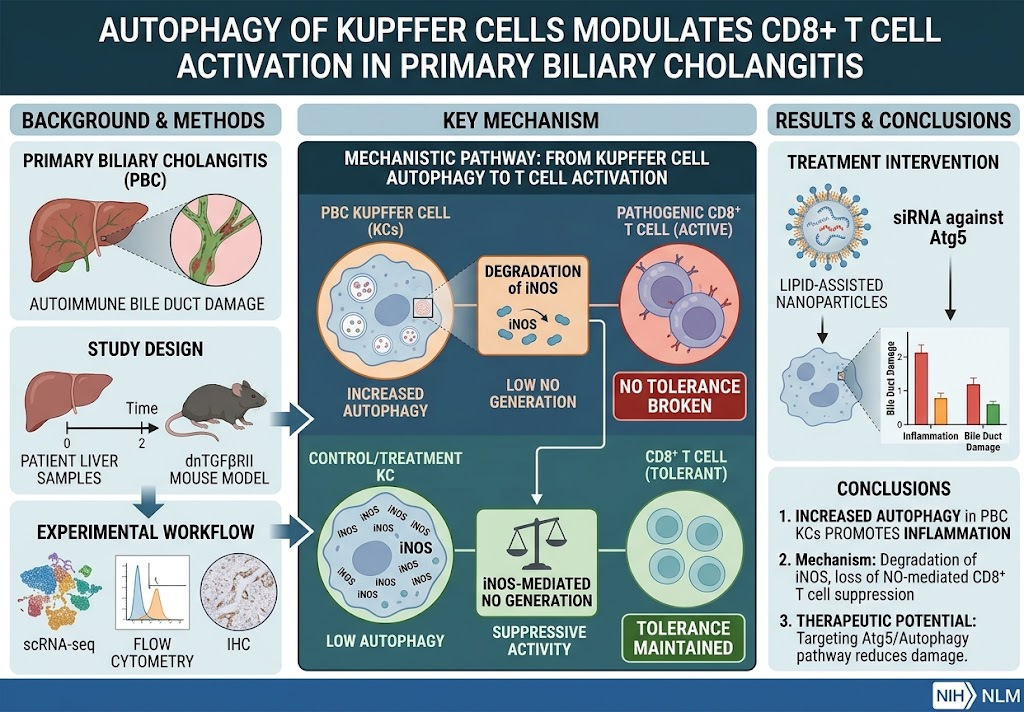
1. Kupffer cells in both human primary biliary cholangitis (PBC) and murine autoimmune cholangitis exhibit increased autophagy and upregulated inflammatory gene expression.
2. Autophagy in Kupffer cells mediates the degradation of inducible nitric oxide synthase (iNOS), impairing their ability to suppress pathogenic CD8+ T cell activation.
3. Macrophage-specific Atg5 knockout in mice decreases liver inflammation and bile duct damage by restoring Kupffer cell-mediated immune tolerance.
4. Therapeutic targeting of Kupffer cell autophagy via siRNA-loaded cationic lipid nanoparticles reduces autoimmune liver injury, offering a novel treatment avenue for PBC.
Study Background
Primary biliary cholangitis is a chronic autoimmune cholestatic liver disease characterized by progressive destruction of intrahepatic bile ducts. This leads to cholestasis, liver inflammation, fibrosis, and eventual liver failure if untreated. Current therapies mitigate disease progression but do not fully address underlying immune dysregulation. The liver’s immune microenvironment, particularly involving hepatic macrophages—including resident Kupffer cells and infiltrating monocyte-derived macrophages (MoMs)—plays a critical role in modulating immune tolerance and inflammation. Understanding how Kupffer cells regulate autoreactive CD8+ T cells, key effectors in PBC pathogenesis, is essential to identify novel therapeutic targets.
Study Design
The study combined human and murine models to interrogate hepatic macrophage dynamics and autophagy mechanisms. It included phenotypic profiling of liver macrophages from patients with PBC and dnTGFβRII transgenic mice (a model of autoimmune cholangitis) using single-cell RNA sequencing, flow cytometry, and immunohistochemistry. Functional studies involved depleting hepatic macrophages or selectively inhibiting monocyte-derived macrophages in mice, alongside Lyz2-Cre-driven Atg5 knockout to disrupt autophagy in macrophages. Coculture experiments assessed the interaction between Kupffer cells and CD8+ T cells with specific focus on immune tolerance mediators. Finally, therapeutic intervention was tested by delivering small interfering RNA (siRNA) against Atg5 encapsulated in cationic lipid-assisted nanoparticles aimed at Kupffer cells in vivo.
Key Findings
Increased Autophagy and Inflammatory Gene Expression in Kupffer Cells: Kupffer cells isolated from both human PBC patients and dnTGFβRII mice demonstrated elevated expression of genes associated with inflammatory pathways and autophagy, notably increased Atg5 which facilitates autophagosome formation. This autophagic activity correlated with disease severity and inflammatory status.
Macrophage Autophagy Modulates CD8+ T Cell Activation: Kupffer cells normally sustain CD8+ T cell tolerance through producing inducible nitric oxide synthase (iNOS), which generates nitric oxide (NO) to suppress T cell activation. However, upregulated autophagy causes selective degradation of iNOS in Kupffer cells. This degradation impairs their suppressive function, allowing pathogenic CD8+ T cells to become activated and exacerbate bile duct injury.
Atg5 Knockout in Macrophages Ameliorates Liver Injury: Murine models with macrophage-specific Atg5 deletion exhibited reduced hepatic inflammation and less bile duct damage. This protective effect was linked to preserved iNOS levels and enhanced Kupffer cell-mediated immune tolerance toward CD8+ T cells.
Nanoparticle-Mediated Atg5 Silencing Offers Therapeutic Benefit: Administration of cationic lipid-based nanoparticles encapsulating siRNA targeting Atg5 selectively downregulated macrophage autophagy in vivo. This intervention significantly lowered liver inflammation and bile duct destruction in autoimmune cholangitis models, demonstrating translational potential for PBC therapy.
Safety and Efficacy: The study did not report significant adverse effects related to macrophage depletion or Atg5 silencing, indicating a targeted approach may mitigate systemic immunosuppression risks while providing disease-modifying benefits.
Expert Commentary
This research presents compelling mechanistic insights into how Kupffer cell autophagy facilitates autoimmune liver injury by undermining immune tolerance. The demonstration that autophagic degradation of iNOS disrupts Kupffer cell regulatory function underscores the complexity of immune cell cross-talk in PBC. The possibility of employing nanotechnology to deliver siRNA therapeutics specifically to hepatic macrophages opens innovative avenues to modulate the hepatic immune microenvironment without global immunosuppression.
While promising, it is important to note potential limitations including the translatability of murine dnTGFβRII model data to human PBC heterogeneity. Additionally, long-term effects and possible compensatory immune pathways upon macrophage autophagy inhibition warrant further investigation before clinical application. Nonetheless, this study provides a strong rationale for clinical exploration of autophagy-targeted therapies in autoimmune cholangitis.
Conclusion
Macrophage autophagy in Kupffer cells plays a pivotal role in promoting CD8+ T cell activation and autoimmune cholangitis pathogenesis. Targeting Atg5-dependent autophagy restores Kupffer cell-mediated immune tolerance by preserving iNOS expression, thereby reducing liver inflammation and bile duct damage. This novel approach using nanoparticle-mediated Atg5 silencing holds great therapeutic promise for PBC, addressing a critical unmet need in managing this chronic autoimmune liver disease. Further clinical studies are warranted to validate safety and efficacy in patients.
Funding and ClinicalTrials.gov
The original study was supported by grants from relevant research institutions as stated in the source publication (Luo et al., Gut, 2026). No clinical trials are registered specifically for this therapeutic approach as of yet.
References
1. Luo PY, Ma M, Liu MC, Wu YH, Long J, Huang MX, Yang SY, Da TT, Tsuneyama K, Li Q, Jin H, Zhao ZB, Wang J, Ma X, Gershwin ME, Han Y, Lian ZX, Li L. Autophagy of Kupffer cells modulates CD8+ T cell activation in primary biliary cholangitis. Gut. 2026 Aug 6;75(9):1831-1844. PMID: 41371935.
2. Lian ZX, Chuang YH, Tsuneyama K, et al. Primary biliary cirrhosis: autoimmune epithelitis. Autoimmun Rev. 2005;4(2):103-107.
3. Wynn TA. Cellular and molecular mechanisms of fibrosis. J Pathol. 2008;214(2):199-210.
4. Jenne CN, Kubes P. Immune surveillance by the liver. Nat Immunol. 2013;14(10):996-1006.
5. Mizushima N, Komatsu M. Autophagy: renovation of cells and tissues. Cell. 2011;147(4):728-741.