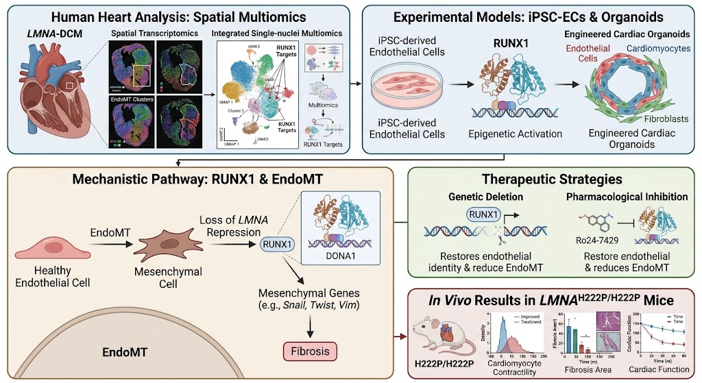
Highlight
• RUNX1 activation drives endothelial-to-mesenchymal transition (EndoMT) in LMNA cardiomyopathy, linking endothelial dysfunction to myocardial fibrosis.
• Loss of LMNA-mediated repression triggers epigenetic and transcriptional reprogramming of endothelial cells, promoting EndoMT.
• Genetic or pharmacological inhibition of RUNX1 restores endothelial identity, reverses EndoMT signatures, and improves cardiac function in cellular and murine LMNA-DCM models.
• Targeting RUNX1 signaling offers a promising therapeutic strategy for fibrotic remodeling in LMNA-associated dilated cardiomyopathy.
Study Background
LMNA-related dilated cardiomyopathy (LMNA-DCM) is a progressive, inherited cardiac disorder caused by mutations in the LMNA gene, which encodes nuclear envelope proteins lamin A and C. Clinically, patients exhibit conduction system disease, life-threatening arrhythmias, myocardial fibrosis, and progressive heart failure, often culminating in heart transplantation or sudden cardiac death. Despite advances, the pathophysiologic mechanisms underlying fibrotic remodeling in LMNA-DCM remain incompletely understood. Historically, LMNA mutations have been linked primarily to intrinsic defects in cardiomyocytes, including nuclear structural abnormalities and gene expression dysregulation. However, the contribution of non-cardiomyocyte compartments, particularly endothelial cells (ECs), to the fibrotic phenotype has not been thoroughly explored.
Endothelial-to-mesenchymal transition (EndoMT) is an emerging mechanism by which endothelial cells lose their characteristic phenotype and acquire mesenchymal, fibroblast-like properties that contribute to fibrotic remodeling in multiple organs, including the heart. The molecular regulators driving EndoMT in LMNA cardiomyopathy have not previously been delineated.
Study Design
This translational study employed comprehensive mechanistic analyses integrating spatial transcriptomics, single-nuclei multiomics, patient-derived cellular models, engineered cardiac organoids, and a genetically engineered LMNA mouse model (LMNAH222P/H222P) to elucidate endothelial transcriptional and epigenomic alterations associated with fibrosis in LMNA-DCM.
The key approaches included:
- Spatial transcriptomics and integrated single-nuclei multiomics analyses of explanted human LMNA-DCM hearts to identify endothelial cell subpopulations with EndoMT-associated signatures.
- Generation of patient-specific induced pluripotent stem cell (iPSC)-derived endothelial cells to study endothelial dysfunction and RUNX1 activation pathways ex vivo.
- Use of engineered multicellular cardiac organoids combining endothelial and cardiomyocyte populations to evaluate functional interactions impacted by endothelial RUNX1 signaling.
- In vivo evaluation of RUNX1’s role through genetic deletion and pharmacologic inhibition (Ro24-7429) in the LMNAH222P/H222P murine model—an established system recapitulating human LMNA-DCM pathology.
- Functional endpoints included endothelial identity restoration, reversal of EndoMT transcriptional programs, chromatin accessibility normalization, cardiomyocyte contractility, myocardial fibrosis quantification, and overall cardiac function.
Key Findings
Identification of RUNX1-driven EndoMT in human LMNA-DCM hearts: Spatial transcriptomic and single-nuclei multiomic profiling of explanted LMNA-DCM hearts revealed distinct endothelial cell populations exhibiting gene expression and chromatin patterns indicative of EndoMT. Notably, these populations demonstrated epigenetic activation and upregulation of RUNX1, a transcription factor previously implicated in mesenchymal transition processes but not extensively studied in LMNA cardiomyopathy.
Mechanistic link between LMNA mutations and endothelial reprogramming: iPSC-derived endothelial cells from patients harboring LMNA mutations exhibited endothelial dysfunction features, activation of mesenchymal genes, and derepression of RUNX1 at both transcriptional and epigenomic levels, implicating LMNA loss as a driver of altered endothelial programming via RUNX1.
Genetic RUNX1 deletion reverses EndoMT and restores endothelial identity: Targeted deletion of RUNX1 in patient-derived endothelial cells normalized endothelial marker expression, reversed mesenchymal gene activation, and restored chromatin accessibility at endothelial regulatory loci, demonstrating a direct causal role for RUNX1 in maintaining the EndoMT phenotype.
Endothelial RUNX1 activation impairs myocardial function in engineered organoids: In multicellular cardiac organoids, activation of RUNX1 in endothelial cells disrupted endothelial-cardiomyocyte crosstalk and decreased cardiomyocyte contractility. Conversely, endothelial-specific RUNX1 deletion rescued these dysfunctions, highlighting the paracrine and mechanistic influence of endothelial RUNX1 on myocardial performance.
Pharmacological RUNX1 inhibition improves cardiac phenotype in LMNA mouse model: Treatment of LMNAH222P/H222P mice with the RUNX1 inhibitor Ro24-7429 after disease onset significantly reduced myocardial fibrosis and preserved cardiac function. These findings establish RUNX1 as a viable therapeutic target and demonstrate the translational potential of modulating endothelial transcriptional reprogramming in fibrotic cardiomyopathy.
Expert Commentary
The study by Wu et al. provides compelling evidence for the role of endothelial RUNX1-driven EndoMT in the pathogenesis of LMNA cardiomyopathy, a paradigm shift from the traditional cardiomyocyte-centric view. By leveraging cutting-edge multiomic technologies combined with human disease modeling and in vivo validation, the work elucidates a novel, targetable pathway contributing to myocardial fibrosis and functional decline.
Limitations include the need to further characterize long-term safety and efficacy of RUNX1 inhibition, as RUNX1 also plays roles in hematopoiesis and other essential biological processes. Additional studies should explore the potential systemic effects of RUNX1-targeted therapies and aim to refine delivery strategies to enhance cardiac specificity.
Furthermore, whether RUNX1-driven EndoMT is implicated in other forms of fibrotic cardiomyopathy remains an open question, highlighting a promising avenue for broader cardiovascular research.
Conclusion
This study identifies RUNX1-mediated endothelial-to-mesenchymal transition as a critical mechanistic link between LMNA mutations and fibrotic remodeling in LMNA-related dilated cardiomyopathy. By demonstrating that endothelial transcriptional reprogramming driven by RUNX1 contributes substantially to myocardial fibrosis and dysfunction, and showing that genetic and pharmacologic RUNX1 inhibition can restore endothelial and myocardial function, these findings pave the way for innovative therapeutic strategies targeting endothelial RUNX1 signaling in fibrotic cardiomyopathies. The translation of RUNX1 inhibitors into clinical testing could potentially transform management of this high-risk genetic cardiomyopathy.
Funding and ClinicalTrials.gov
The original study was supported by research grants from academic and governmental sources as detailed by Wu et al. The pharmacologic agent Ro24-7429 was provided by collaborating pharmaceutical entities. No registered clinical trials have yet been reported for RUNX1 inhibitors in LMNA cardiomyopathy, underscoring the novelty and translational potential of the therapeutic approach.
References
- Wu D, Tripathi D, Manhas A, et al. RUNX1-driven endothelial-to-mesenchymal transition contributes to remodelling in LMNA cardiomyopathy. Eur Heart J. 2026 Aug 11; PMID: 42578927.
- Bisol et al. Role of Endothelial-to-Mesenchymal Transition in Cardiac Fibrosis. Front Cardiovasc Med. 2021;8:650024.
- Muchir A, Worman HJ. LMNA-Related Cardiomyopathies. Circ Res. 2021;128(4):567-577.
- Markwald RR, et al. Endothelial to Mesenchymal Transition: A Conduit for Cardiac Fibrosis Associated with Heart Disease. Front Cardiovasc Med. 2022;9:842067.