Highlight
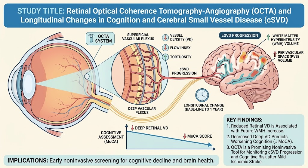
1. Longitudinal decrease in retinal vessel density (VD) assessed by optical coherence tomography angiography (OCTA) correlates with white matter hyperintensity (WMH) progression in cerebral small vessel disease (cSVD).
2. Changes in retinal vascular parameters also associate with increases in basal ganglia perivascular space volume and cognitive decline after mild ischemic stroke.
3. Baseline OCTA measures predict one-year brain imaging and cognitive outcomes, supporting OCTA’s potential as a noninvasive monitoring tool in stroke populations enriched for cSVD.
Study Background
Cerebral small vessel disease (cSVD) is a prevalent cause of vascular cognitive impairment and stroke, contributing significantly to morbidity worldwide. Despite advances in neuroimaging markers such as white matter hyperintensities (WMHs) and perivascular spaces (PVS) on MRI, there remains a need for accessible, noninvasive biomarkers to detect and monitor microvascular pathology longitudinally. The retina provides a unique window into cerebral microcirculation due to its embryologic and anatomical similarities with cerebral small vessels. Optical coherence tomography angiography (OCTA) enables detailed, dye-free visualization and quantification of retinal microvasculature, including vessel density (VD) and flow characteristics, offering promise for early detection and monitoring of cerebral vascular changes.
Previous cross-sectional studies have suggested associations between retinal microvascular abnormalities detected by OCTA and both cSVD burden and cognitive impairment. However, the longitudinal relationship between retinal vascular changes post-stroke and subsequent brain and cognitive outcomes remains unclear. This study aimed to prospectively characterize retinal microvascular changes using OCTA in relation to MRI markers of cSVD and cognition over one year following mild ischemic stroke, evaluating whether baseline OCTA parameters predict future deterioration.
Study Design
This investigation was a prospective, longitudinal cohort study conducted at a tertiary hospital stroke service. Patients with recent lacunar or mild cortical ischemic stroke (modified Rankin Scale ≤2) were recruited. Assessments were performed at baseline (post-stroke) and at one-year follow-up and included detailed clinical evaluations, cognitive testing via the Montreal Cognitive Assessment (MoCA), brain MRI, and retinal imaging using OCTA.
OCTA metrics comprised vessel density (VD), foveal avascular zone (FAZ) area, vessel radius, tortuosity, and flow index from both superficial and deep retinal vascular layers. Brain MRI outcomes focused on quantifiable markers of cSVD, specifically the volume of WMHs, basal ganglia perivascular spaces (PVS), and mean diffusivity within normal-appearing white matter. The analysis employed linear mixed-effects models to assess (1) longitudinal associations between retinal microvascular changes and brain/cognitive markers and (2) predictive value of baseline retinal measures for one-year brain and cognitive outcomes. Analyses adjusted for confounders including age, sex, systolic blood pressure, smoking, diabetes, stroke severity, disability, and stroke subtype.
Key Findings
The study enrolled 189 participants with mean age 64.8 years (37% female) at baseline; 154 were followed at one year. Key longitudinal findings were:
- Reductions in retinal vessel density (VD) over time in both superficial and deep vascular layers significantly correlated with an increase in WMH volume, indicating progressive microvascular damage associated with retinal changes. Standardized beta coefficients were -0.045 (95% CI: -0.067 to -0.023) for the superficial layer and -0.050 (-0.074 to -0.026) for the deep layer.
- Increasing retinal VD alongside reductions in retinal flow index were linked with greater basal ganglia PVS volume, suggesting complementary microvascular alterations in retinal and cerebral small vessels.
- Declines in MoCA scores over one year were associated with decreases in deep retinal VD; a positive standardized beta of 0.136 (0.036–0.236) supported a link between retinal microvascular integrity and cognitive function.
Moreover, baseline OCTA metrics demonstrated prognostic value:
- Lower baseline VD in both superficial and deep layers predicted worse cognitive performance at one year (superficial: 0.125 [0.043–0.207]; deep: 0.242 [0.162–0.322]).
These findings underscore the utility of OCTA retinal biomarkers as reflections of cerebral microvascular pathology progression and cognitive trajectory following mild ischemic stroke.
Expert Commentary
The study by Gibbon et al. advances the field of vascular neurology and neuro-ophthalmology by longitudinally validating retinal OCTA parameters as biomarkers correlated with cerebral small vessel disease progression and cognitive decline. The strength of the study lies in its prospective design, integration of multimodal imaging, robust adjustment for vascular risk factors, and the inclusion of mild stroke patients who represent a population at risk yet often under-monitored.
Biologically, the retina’s microcirculation shares developmental origins, endothelial characteristics, and autoregulatory mechanisms with cerebral small vessels, supporting the plausibility of parallel vascular changes detectable by OCTA. The association between reduced vessel density and increased WMH volume suggests that capillary rarefaction and hypoperfusion may drive cerebral white matter damage and cognitive dysfunction.
Limitations include the moderate sample size and potential selection bias of mild stroke survivors, which may limit generalizability to more disabled or non-stroke populations. Technological variability and segmentation differences in OCTA warrant standardization before widespread clinical application. Future larger-scale studies are needed to refine retinal OCTA thresholds to stratify risk and evaluate interventions targeting microvascular health.
Conclusion
This longitudinal cohort study establishes that retinal microvascular changes measured by OCTA are closely associated with the progression of cerebral small vessel disease and cognitive decline following mild ischemic stroke. Baseline OCTA retinal vessel density serves as a predictive biomarker of future brain MRI and cognitive outcomes, supporting the integration of retinal imaging into stroke patient management. As a rapid, noninvasive tool, OCTA holds promise for monitoring microvascular pathology and potentially guiding therapeutic strategies in cSVD-related cognitive impairment and stroke.
Further research should focus on validating these findings in larger, diverse cohorts, exploring mechanistic pathways linking retinal and cerebral microvascular disease, and assessing the impact of retinal biomarker-guided interventions to improve cerebrovascular health and functional outcomes.
Funding and Clinical Trials
The study was funded by the Stroke Association [specific grant numbers not detailed]. No clinical trial registration was indicated for this observational cohort study.
References
1. Gibbon S, Giarratano Y, Hamid C, et al. Retinal Optical Coherence Tomography-Angiography and Longitudinal Changes in Cognition and Cerebral Small Vessel Disease. Neurology. 2026 Sep 15;107(7):e2185-02. PMID: 42743445.
2. Wardlaw JM, Smith C, Dichgans M. Small vessel disease: mechanisms and clinical implications. Lancet Neurol. 2019 Jul;18(7):684–696.
3. McGrory S, Wardlaw JM. Cerebral small vessel disease: the importance of recognizing the multifactorial nature of this brain disease. Hypertension. 2021;77(1):3–5.
4. Shi Y, Wardlaw JM. Update on cerebral small vessel disease: a dynamic whole-brain disease. Stroke Vasc Neurol. 2016 Sep;1(3):83-92.
5. Zhong R, Zhang Z, Jin L, et al. Retinal microvasculature and brain small vessel disease: a cross-sectional study using optical coherence tomography angiography. Stroke. 2021;52(3):e9–11.