
Highlight
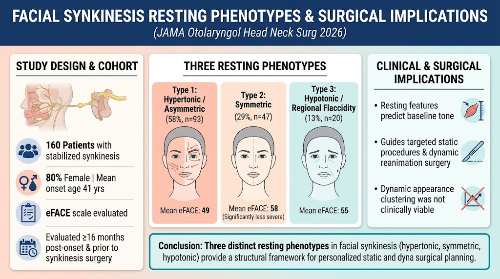
This cross-sectional analysis of 160 patients with stabilized facial synkinesis identified three clinically meaningful resting phenotypes—type 1 characterized by regional hypertonicity and asymmetry, type 2 by facial symmetry with less severe synkinesis, and type 3 by localized hypotonicity. Recognition of these patterns offers an evidence-based framework to tailor static and dynamic surgical interventions.
Study Background
Facial synkinesis is a challenging complication following facial nerve paralysis, characterized by involuntary muscle contractions during voluntary movements. The condition creates both functional impairment and psychosocial distress due to facial asymmetry and abnormal expressions. Despite advances in treatment options like targeted chemodenervation and surgical reconstruction, there has been a lack of phenotypic stratification based on resting facial features to guide personalized management strategies. This study addresses the unmet need by classifying phenotypes of facial synkinesis at rest, which may influence decision-making for static and dynamic corrective procedures.
Study Design
This retrospective cross-sectional study was conducted at a tertiary academic referral center, analyzing videographic and photographic standardized data collected between January 2009 and March 2025. Eligible patients presented with unilateral facial synkinesis stabilized at least 16 months post-onset of paralysis. Inclusion required availability of standardized resting facial photographs and videos prior to any synkinesis-focused surgeries and beyond three months after last chemodenervation treatments to prevent treatment interference with phenotype assessment. Patients with bilateral synkinesis or missing required images were excluded. Evaluations were made independently by three expert raters using the clinician-graded eFACE facial nerve scale to quantify synkinesis severity. A clustering analytical approach was applied to identify resting facial phenotypes.
Key Findings
Among 549 consecutive facial synkinesis patients reviewed, 160 met criteria for inclusion. This cohort had a predominance of females (80%) with a mean age of 41 years at initial paralysis. The median follow-up time was 1.1 years after paralysis onset, with a median interval to evaluation of 5.4 years. The average eFACE synkinesis score was 53 out of 100, signifying moderate synkinesis severity.
Three distinct phenotypic clusters emerged at rest:
- Type 1 (58%): This most common phenotype showed significant regional hypertonicity and resting facial asymmetry, reflected by a mean eFACE score of 49, indicating more severe synkinesis. These patients often have marked involuntary muscle tightness causing notable asymmetry at rest.
- Type 2 (29%): Patients had notable facial symmetry at rest with less severe synkinesis (mean eFACE score 58). This group showed less resting muscle hypertonicity, suggesting potential for better baseline facial tone but dynamic impairments during movement.
- Type 3 (13%): Characterized by localized hypotonicity on the affected side and moderate synkinesis severity (mean eFACE 55), this less common phenotype may represent atypical muscle weakness rather than hypertonic synkinetic contractions.
Attempts to define clusters based on dynamic facial appearances did not produce clinically useful stratifications.
Importantly, the eFACE synkinesis score differed significantly between type 1 and type 2 phenotypes, supporting that resting hypertonicity associates with more severe synkinesis manifestations.
Expert Commentary
This study contributes a critical step forward in conceptualizing facial synkinesis, moving beyond symptom recognition to phenotype-driven clinical stratification. By delineating resting hypertonic, symmetric, and hypotonic patterns, clinicians can better anticipate functional deficits and tailor interventions.
Type 1 patients with regional hypertonicity might benefit from combination approaches focusing on chemodenervation to reduce involuntary muscle contractions alongside surgical releases or muscle transfers to restore symmetry and function. Conversely, type 2 patients’ symmetrical resting tone but residual synkinesis may call for dynamic neuromuscular retraining and selective chemodenervation. Type 3’s hypotonicity implies a need for muscle strengthening or augmentation strategies rather than tone reduction.
Currently, treatment decisions often rely on subjective assessments. Incorporating this phenotypic framework could standardize evaluation, enable prognostication, and optimize surgical planning.
Limitations include its retrospective design and single-center data, which may limit generalizability. Also, the lack of a dynamic phenotype is an area for future investigation with larger patient datasets or advanced motion analysis techniques.
Conclusion
This investigation of resting facial synkinesis phenotypes identifies three clinically meaningful clusters with distinctive features of hypertonicity, symmetry, and hypotonicity. Recognition of these phenotypes provides a systematic evaluative tool that can refine surgical treatment planning, integrating static and dynamic strategies to improve outcomes. Future prospective studies validating this classification and linking phenotypes with long-term treatment responses are warranted.
Funding and ClinicalTrials.gov
Details regarding funding and clinical trial registration were not provided in the available abstract.
References
- Bhatia SS, Johansen CF, Patel DR, et al. Facial Synkinesis Resting Phenotypes and Surgical Implications. JAMA Otolaryngol Head Neck Surg. 2026 Oct 15. PMID: 42879147.
- Manktelow RT, Zuker RM, Hadlock TA. Surgical rehabilitation of facial paralysis with regional muscle transfer. Clin Plast Surg. 2016;43(3):381-397.
- Hadlock TA. Facial nerve paralysis and synkinesis: Electrophysiology and treatment strategies. Facial Plast Surg. 2014;30(3):199-205.
- Chen KS, Grassmann JP, Myckatyn TM. Quantifying facial synkinesis and outcomes after botulinum toxin treatment: A comprehensive review. Otolaryngol Head Neck Surg. 2020;162(6):760-768.