Prior soft-tissue augmentation with autogenous subepithelial connective tissue graft (SCTG) before flapless implant placement significantly enhances peri-implant soft-tissue dimensions compared to simultaneous augmentation or no augmentation.
Both autogenous grafts and collagen-based substitutes contribute to soft tissue volume gains, but autogenous grafts outperform collagen substitutes in increasing lingual peri-implant tissue thickness.
All groups experienced crestal bone remodeling around implants, indicating soft-tissue augmentation does not prevent physiological bone changes post-implantation.
The study supports the potential benefits of staged augmentation strategies to optimize soft tissue outcomes in implant dentistry, warranting confirmation by clinical trials.
Study Background
Dental implant therapy has revolutionized oral rehabilitation, yet the maintenance of peri-implant soft tissue dimensions remains critical to long-term implant success. Adequate soft tissue volume around implants improves esthetic outcomes, tissue stability, and may reduce peri-implantitis risk. Various grafting materials and timing protocols for crestal soft-tissue augmentation have been proposed to enhance peri-implant tissue dimensions. However, robust evidence comparing the histological impact of augmenting soft tissue either prior to or simultaneously with implant placement, using autogenous connective tissue grafts versus collagen-based substitutes, remains limited. This study addresses this gap by providing controlled experimental in vivo data to inform optimal surgical timing and graft materials to maximize peri-implant tissue thickness.
Study Design
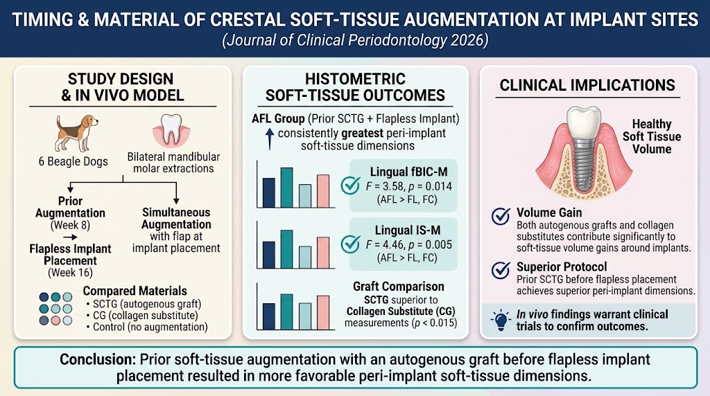
This preclinical study employed a controlled, split-mouth experimental design in six beagle dogs. Bilateral extractions of mandibular first, second, and third molars were performed to create edentulous sites.
Eight weeks post-extraction, one mandibular side underwent primary crestal soft-tissue augmentation with either an autogenous subepithelial connective tissue graft (SCTG), a collagen-based graft substitute (CG x1D), or no augmentation (control). At Week 16 (eight weeks after augmentation), implants were placed flaplessly at these sites without additional grafting.
Conversely, on the contralateral side, implant placement with flap elevation was performed simultaneously with either SCTG, CG x1D, or no augmentation at Week 16 without prior soft-tissue augmentation. Animals were euthanized at Week 36 for histological analysis.
Histometric endpoints included quantitative measures of crestal bone remodeling and peri-implant soft-tissue dimensions, such as the distance from the first bone-to-implant contact to the implant shoulder (fBIC-M) and implant shoulder to soft tissue margin (IS-M). Data were analyzed using linear mixed-effects models to account for intra-animal variability, followed by post-hoc Tukey adjustments for group comparisons.
Key Findings
All experimental groups exhibited expected crestal bone remodeling apically to the implant shoulder, reflecting physiological bone adaptation after implant placement. No grafting strategy prevented this remodeling.
Critically, the group receiving autogenous SCTG augmentation prior to flapless implant placement (denoted AFL) demonstrated significantly greater peri-implant soft-tissue dimensions compared to groups receiving simultaneous augmentation or no augmentation. Specifically, the AFL group showed increased lingual site thickness measured by fBIC-M (F=3.58, p=0.014) and IS-M (F=4.46, p=0.005), exceeding values in the simultaneous flap elevation (FL) and control (FC) groups.
Further analyses revealed that autogenous SCTG sites outperformed collagen substitute sites in enhancing lingual soft-tissue thickness, with significant differences in fBIC-M (p=0.013) and IS-M (p=0.012). This highlights the superior volumetric stability and tissue integration capacity of autogenous connective tissue grafts over collagen-based materials in this setting.
Importantly, both graft materials yielded soft-tissue volume gains relative to no augmentation, suggesting that collagen substitutes remain viable alternatives when autogenous graft availability or patient morbidity is a concern.
Safety and Observations
No adverse effects or complications such as infection, excessive bone loss, or graft exposure were reported during the study period, supporting the safety of both augmentation timings and graft materials under controlled conditions.
Expert Commentary
This comprehensive in vivo study provides histological evidence favoring a staged surgical approach, where soft-tissue augmentation with autogenous grafts precedes implant placement, optimizing peri-implant tissue dimensions that are essential for esthetics and long-term stability. The flapless implant placement following prior augmentation likely preserves the augmented tissue volume better than simultaneous flap elevation procedures.
Despite the promising findings, translation to human clinical practice requires caution due to interspecies differences and controlled experimental conditions. Additionally, the sample size of six dogs, while reasonable in preclinical research, limits generalizability. Future randomized clinical trials with larger patient cohorts and longer follow-up are needed to validate these preclinical outcomes and assess clinical parameters such as patient morbidity, esthetic satisfaction, and implant survival.
The study also underscores the plausible biological advantage of autogenous connective tissue grafts, which contain viable fibroblasts and extracellular matrix components critical for vascularization and volume maintenance. Collagen substitutes offer practical benefits but may not replicate the full biological milieu necessary for maximal tissue thickening.
Conclusion
Prior crestal soft-tissue augmentation using autogenous subepithelial connective tissue grafts before flapless implant placement significantly enhances peri-implant soft-tissue dimensions compared with simultaneous augmentation or no augmentation. Collagen-based graft substitutes also contribute to volume gain but are inferior to autogenous grafts in this model. These findings suggest that a staged augmentation approach may provide superior peri-implant tissue quality and volume, potentially improving clinical outcomes in implant dentistry. Well-designed clinical trials are warranted to confirm these results and guide evidence-based surgical protocols.
Funding and ClinicalTrials.gov
No specific funding or clinical trial registration information was provided by the authors.
References
1. Zhang Y, Park SY, Yoo SH, Bae SL, Pachiou A, Jung UW, Thoma DS. Effect of Crestal Soft-Tissue Augmentation Prior to or Simultaneously With Implant Placement on Peri-Implant Tissue Dimensions: Histological Outcomes of an Experimental In Vivo Study. J Clin Periodontol. 2026 Oct 11. PMID: 42860263.
2. Thoma DS, et al. Soft tissue volume augmentation at dental implant sites: a systematic review. Clin Oral Implants Res. 2018;29 Suppl 15:269-281.
3. Gränicher P, et al. Peri-implant soft tissue stability and keratinized mucosa width around implants placed with immediate and early protocols – A review. Clin Oral Implants Res. 2020;31(1):24-39.
4. Cairo F, et al. Soft tissues around dental implants: biological and clinical considerations. J Appl Oral Sci. 2018;26:e20170439.