Highlight
- Paramagnetic rim lesions (PRLs) on susceptibility-sensitive MRI identify chronic active inflammatory lesions in multiple sclerosis (MS).
- The presence of PRLs is associated with increased risk of disability progression independent of relapse activity (PIRA) over 5 years.
- PRL burden correlates with future rates of brain atrophy and markers of chronic intrathecal inflammation.
- PRLs hold promise as non-invasive prognostic biomarkers aiding individual risk stratification and therapeutic decisions in MS management.
Study Background
Multiple sclerosis is a chronic autoimmune-mediated neurological disorder characterized by relapsing inflammatory attacks and progressive neurodegeneration. While relapse events contribute to clinical disability, accumulating evidence emphasizes progression independent of relapse activity (PIRA) as a major driver of long-term disability accumulation. Identifying reliable biomarkers capable of predicting PIRA and neurodegeneration remains a high priority to optimize personalized treatment approaches.
Paramagnetic rim lesions (PRLs) represent a novel radiologic marker visualized on susceptibility-weighted or quantitative susceptibility mapping (QSM) MRI sequences. They correspond histopathologically to iron-laden microglia/macrophages encapsulating chronic active lesions, reflecting ongoing inflammation and tissue injury. Initial studies have linked PRLs to greater disability and worse clinical outcomes, but robust longitudinal data defining their prognostic value for PIRA and brain atrophy over extended follow-up are limited.
Study Design
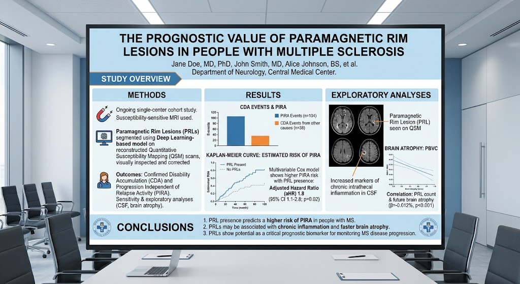
This investigation utilized a large, ongoing single-center cohort of people with MS who underwent susceptibility-sensitive MRI allowing assessment of PRLs via QSM. Using a deep learning segmentation model followed by expert review, PRLs were identified and quantified. The cohort included 862 participants (mean age 40.3 years; 64.7% female). Clinical follow-up data spanning a median of 5.1 years enabled analysis of outcomes including confirmed disability accumulation (CDA) and PIRA events.
Multivariable Cox proportional hazards models were applied to assess the prognostic significance of PRL presence for disability progression, adjusting for relevant covariates. Sensitivity analyses explored the impact of PRL count, MS subtype, prior disability status, and treatment during follow-up. Additionally, exploratory analyses evaluated associations between PRL status, cerebrospinal fluid (CSF) biomarkers indicative of chronic inflammation, and annualized brain volume changes as markers of neurodegeneration.
Key Findings
Among 862 MS patients, 395 (45.8%) exhibited at least one PRL on MRI. Within the subset of 801 individuals with longitudinal clinical data, 142 (17.7%) experienced CDA over follow-up, with 104 of these CDA events (73.2%) classified as progression independent of relapse activity (PIRA).
Critically, the presence of PRLs was associated with a significantly increased risk of developing PIRA (adjusted hazard ratio 1.8; 95% confidence interval [CI], 1.1–2.8; p = 0.02), indicating nearly double the likelihood of progression independent of relapses compared to those without PRLs.
Exploratory subgroup analyses revealed that individuals harboring PRLs had higher levels of CSF markers associated with chronic intrathecal inflammation, such as elevated immunoglobulin indices and oligoclonal bands, supporting the biological plausibility of PRLs reflecting persistent inflammatory activity.
Moreover, a dose-dependent relationship was observed between the number of PRLs and the rate of brain atrophy measured by annual percentage brain volume change (PBVC) (β = -0.012%, 95% CI -0.018% to -0.005%; p < 0.001), indicating greater structural neurodegeneration correlating with greater PRL burden.
These findings were robust across sensitivity analyses accounting for different MS phenotypes, previous disability accumulation, and disease-modifying therapy exposure, suggesting PRLs hold independent prognostic value.
Expert Commentary
This study substantially advances understanding of PRLs as imaging biomarkers by quantifying their prognostic significance for clinically meaningful progression independent of relapse in MS. The demonstration of a nearly twofold increased risk for PIRA and correlation with future brain atrophy underscores the potential clinical utility of incorporating PRL assessment in routine MRI protocols.
Importantly, PRLs reflect ongoing chronic lesion activity driven by iron-laden inflammatory cells, offering a mechanistic explanation for their association with sustained tissue injury and neurodegeneration beyond acute inflammatory relapses. This biological insight bolsters confidence in PRLs as a surrogate marker capturing a distinct and clinically relevant disease process.
Limitations include the single-center design, which may limit generalizability, and the lack of standardized MRI acquisition protocols globally. Future multicenter studies with harmonized imaging methods are needed to validate these findings and facilitate translation into clinical practice.
Additionally, longitudinal interventions targeting PRLs specifically remain unexplored, representing an avenue for clinical trials. Incorporating PRLs into predictive algorithms alongside clinical and fluid biomarkers could refine risk stratification and guide individualized therapeutic escalation, particularly for progressive MS phenotypes.
Conclusion
This longitudinal cohort study highlights paramagnetic rim lesions as independent predictors of progression independent of relapse activity and accelerated brain atrophy in multiple sclerosis. PRLs serve as accessible, non-invasive MRI biomarkers that reflect chronic lesion activity and underlying inflammation driving neurodegeneration. Their integration into clinical assessment may enable earlier identification of patients at elevated risk for disability progression, thereby informing personalized management and improving long-term outcomes.
Further research is needed to standardize PRL imaging protocols, validate prognostic models in diverse populations, and evaluate targeted treatment strategies addressing chronic active lesions in MS.
Reference
Lauerer M, Voon CC, McGinnis J, Sepp D, Meineke J, Wiltgen T, Berg RC, Preibisch C, Wiestler B, Engl C, Uibel P, Berthele A, Kirschke JS, Hemmer B, Mühlau M. The Prognostic Value of Paramagnetic Rim Lesions in People With Multiple Sclerosis. Neurology. 2026 Sep 22;107(6):e218428. doi: 10.1212/WNL.0000000000218428. Epub 2026 Aug 20. PMID: 42623573.
Granberg T. Paramagnetic Rim Lesions and Their Predictive Value of Future Progression in People With Multiple Sclerosis. Neurology. 2026 Sep 22;107(6):e218582. doi: 10.1212/WNL.0000000000218582. Epub 2026 Aug 20. PMID: 42623576.