Introduction and Context
FLT3-internal tandem duplication (FLT3‑ITD) is one of the most important somatic lesions in acute myeloid leukemia (AML). FLT3‑ITD status at diagnosis informs prognosis and therapy selection (for example, FLT3 inhibitors). Until recently, FLT3‑ITD measurable residual disease (MRD) assessment was technically challenging: heterogeneous insertion lengths, variable insertion sites, and PCR/sequencing bias limited assay sensitivity and reproducibility. As next‑generation sequencing (NGS) methods matured — including unique molecular identifiers (UMIs), targeted hybrid capture, and robust bioinformatics — reliable, sensitive FLT3‑ITD detection at MRD-relevant levels became feasible.
The ELN‑DAVID (European LeukemiaNet — Diagnostic and Validation of IDentification) recommendations published in Blood (Hourigan et al., 2026) provide a focused, practical, consensus technical and clinical framework for NGS-based FLT3‑ITD MRD testing. This companion technical note aligns laboratory standards, reporting elements, timing of tests, and clinical interpretation to support use of FLT3‑ITD MRD in routine practice and trials.
Why this guidance now: key drivers
– Technical progress: UMIs, bespoke informatics and improved library methods substantially lower error rates and recover variable-length ITDs.
– Clinical need: FLT3 inhibitors (midostaurin, gilteritinib, quizartinib in trials) and transplantation decisions create scenarios where robust MRD data on FLT3‑ITD can change management.
– Prior gap: earlier ELN statements acknowledged FLT3‑ITD as prognostic but did not recommend routine MRD testing because of assay limitations. The new ELN‑DAVID consensus closes that gap with lab and clinical recommendations to standardize practice.
New Guideline Highlights
Major practical takeaways
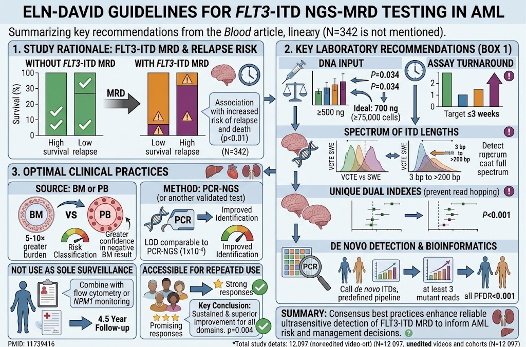
– ELN‑DAVID endorses validated NGS assays for FLT3‑ITD MRD testing in clinical care and trials when laboratories meet performance and reporting standards.
– Bone marrow (BM) is preferred for MRD sensitivity; peripheral blood (PB) is acceptable for serial surveillance where validated and when bone marrow is not feasible.
– Recommended analytical sensitivity (limit of detection, LOD): assays should reliably detect FLT3‑ITD at or below roughly 10^‑4 variant allele fraction (VAF; 0.01%), with documented performance; best‑practice labs aim for 10^‑5 where validated.
– Use of UMIs and orthogonal validation (replicate libraries, spike‑in controls) is recommended to control PCR/sequencing artifacts and overcome length bias.
– Standardized reporting elements are required (see below), including ITD sequence, insertion site, length, allelic ratio at diagnosis and MRD timepoints, assay sensitivity, and interpretive comment.
– FLT3‑ITD MRD positivity should be interpreted in the clinical context (co‑mutations, prior allelic ratio, therapy), and used to guide discussions around preemptive therapy and transplant stratification rather than as a sole determinant.
Updated Recommendations and Key Changes from Previous Guidance
Summary of change vs prior ELN statements
– Previous ELN guidance (pre‑2026) recognized FLT3‑ITD’s prognostic importance but did not provide standardized MRD testing guidance because of technical variability. ELN‑DAVID moves from “not routinely recommended” to “recommended when performed with validated NGS methods.”
– New explicit technical expectations: LOD, use of UMIs, sample type preference, and bioinformatics validation requirements are now specified, whereas prior documents left these to individual labs.
– Standardized reporting template: ELN‑DAVID requires minimum reporting fields to improve cross‑center comparability — an important upgrade.
Why evidence supports the update
Prospective and retrospective studies over the prior decade increasingly show that persistence or re‑emergence of FLT3‑ITD after initial therapy predicts relapse, and that FLT3‑ITD allele burden dynamics correlate with outcomes. The availability of actionable FLT3 inhibitors and strategies to direct transplant timing/conditioning provide clinical leverage for MRD information.
Topic-by-Topic Recommendations
Laboratory and assay design (technical requirements)
– Assay architecture: targeted NGS panels specifically designed to recover ITDs are recommended, using strategies that span the juxtamembrane / TKD boundary and accommodate insertion heterogeneity. PCR‑bias reducing approaches (hybrid capture, anchored multiplex PCR) are acceptable when validated.
– UMIs (molecular barcodes): recommended to enable error correction and accurate low VAF calls.
– Analytical performance: labs must validate and document LOD, limit of quantification (LOQ), linearity, precision, specificity, and accuracy. Routine target: LOD ≤ 1 × 10^‑4 VAF; aspiration: ≤ 1 × 10^‑5.
– Controls: include contrived samples or characterized clinical specimens with known ITD sizes and VAFs; negative controls; and periodic proficiency testing.
– Bioinformatics: pipelines should be validated to call variable-length indels, provide sequence-resolved calls (exact inserted sequence and position), and quantify allelic ratio. Reporting must note the pipeline version and validated performance characteristics.
Sample type and timing
– Sample choice: bone marrow aspirate is preferred for initial MRD assessment post‑induction and pre‑transplant due to superior sensitivity; peripheral blood is acceptable for frequent surveillance if the local validation demonstrates comparable sensitivity and concordance.
– Recommended testing timepoints (minimum):
– End of induction (to document deep remission or persistence)
– End of consolidation (to document further clearance)
– Pre‑allogeneic stem cell transplant (to inform conditioning and risk assessment)
– Scheduled surveillance every 1–3 months in the first year (PB or BM per local validation) and clinically driven testing thereafter
– At any clinical suspicion of relapse
Reporting standards (required elements)
Every clinical report should include:
– Specimen type and collection date
– Assay method and version (library method, use of UMIs, sequencing platform)
– LOD/LOQ for that run
– FLT3‑ITD identification: exact inserted sequence (where feasible), insertion site (exon/intron), length of ITD
– Allelic ratio or VAF at diagnosis and each MRD timepoint; if assay quantitation below LOQ, report as “detected below quantification limit” with LOD stated
– Interpretive comment: clinical significance, assay limitations, concordance with co‑mutations, and recommended clinical actions (e.g., confirm in orthogonal assay, intensify surveillance, consider preemptive therapy)
Clinical interpretation and use
– Definition of MRD positivity: detection of the diagnostic FLT3‑ITD clone above the assay LOD is considered MRD‑positive; quantification provides additional prognostic information.
– Risk stratification: persistent FLT3‑ITD post‑treatment is associated with higher relapse risk; higher residual allele burden portends worse outcomes. Interpretation should consider co‑occurring mutations (e.g., NPM1) and clinical state.
– Use to guide therapy: MRD positivity can inform discussions about:
– Allogeneic transplant candidacy and timing (higher relapse risk supports proceeding to transplant)
– Preemptive or maintenance therapy (FLT3 inhibitor maintenance post‑transplant for MRD‑positive patients is an area of active study and may be considered in selected cases)
– Enrollment in MRD‑directed trials
– Caveat: the guideline emphasizes that MRD results should not be the sole trigger for high‑risk interventions without corroboration (clinical context, confirmatory testing, and multidisciplinary discussion are recommended).
Special populations and considerations
– Patients treated with FLT3 inhibitors: therapy can suppress FLT3‑ITD burden and, in some cases, select for resistant clones; serial MRD testing may detect emerging resistance. The report should note prior FLT3 inhibitor exposure.
– Low‑burden clones and clonal hematopoiesis: distinguishing persistent leukemia clones from clonal hematopoiesis or CHIP requires context (co‑mutations, VAF dynamics, and morphology).
– Pediatric AML: principles apply but validation in pediatric cohorts is recommended.
Quality assurance and external validation
– Participation in external quality assessment (EQA) or proficiency testing schemes is recommended as they become available for FLT3‑ITD MRD.
– Internal quality controls and periodic revalidation after major pipeline changes (wet lab or bioinformatics) are mandatory.
Expert Commentary and Insights
Committee perspective
Hourigan and the ELN‑DAVID working group emphasize pragmatic balance: routine FLT3‑ITD MRD testing is now technically possible and clinically meaningful, but only when laboratories meet strict validation and reporting standards. They stress the need for multidisciplinary integration: hematologists, laboratory directors, molecular pathologists, and transplant teams must interpret results together.
Key controversies highlighted by the panel
– Optimal LOD: while ELN‑DAVID sets a recommended target LOD of ~10^‑4, some centers push to 10^‑5. The panel did not mandate a single number, recognizing resource and platform variability.
– PB vs BM for surveillance: PB is more convenient, but sensitivity tradeoffs exist. The group recommends local validation rather than blanket substitution.
– Actionability thresholds: what VAF should trigger intervention? The panel refrains from prescribing a universal cutoff and recommends contextualized decisions until prospective trials define thresholds tied to interventions.
Research and evidence gaps the panel identified
– Prospective trials: randomized trials testing MRD-guided preemptive therapy or transplant strategies are needed to confirm that MRD-driven interventions improve survival.
– Standardized external proficiency programs: to support inter‑laboratory comparability.
– Data on integration with other MRD markers (eg, NPM1 qPCR, flow cytometry) to develop composite MRD algorithms.
Recent evidence of prognostic association between FLT3-ITD detection in remission and clinical outcome of intensively treated patients with AML (with or without FLT3 inhibitors)
| Cohort | Patients, n | Remission | Collection time | Sample | Test | DNA used (ng) |
|---|---|---|---|---|---|---|
| PreMEASURE2 (111 sites CIBMTR, 2013-2019) | 608 | CR1 | CR1 pre-alloHCT (≤100 d) | PB | Custom/Archer | 500 |
| ReMEASURE6 (subset of PreMEASURE) | 537 | CR1 | CR1 pre-alloHCT (≤100 d) | PB | Invivoscribe | 700 |
| UK-Aus Retrospective4 MRC AML17 (2009-2014) Alfred/Royal Melbourne Hospital/Peter Mac (2010-2020) |
104 | CR1 (n = 89),CR2 (n = 15) | CR pre-alloHCT (median, 27.5 d before transplantation; range, 1-87) | BM (n = 92),PB (n = 12) | Modified getITD | 500 |
| HOVON-SAKK3 (multicenter prospective phase 3 trials) | 161 | CR1 | Diagnosis, CR1 (pre-alloHCT, if received transplantation) | BM (n = 156),PB (n = 5) | Exon 14 PCR-NGS | 100 and 500 |
| QuANTUM-First8,12 (phase 3 RCT quizartinib vs placebo) | 308 | Composite CR1 | Time of response assessment during induction in composite CR1 | BM | Exon 14-15 PCR-NGS | — |
| BMT-CTN 15065,13 (MORPHO, phase 3 RCT gilteritinib vs placebo after alloHCT) | 356 | CR1 | CR1 pre-alloHCT, prerandomization, and at 3, 6, 12, 18, and 24 mo after randomization | BM | Invivoscribe | 700 |
| ALLG AMLM1610 (phase 2 RCT of sorafenib vs placebo added to intensive induction) | 74 | CR1 | CR/CRi after induction | BM | Modified getITD | 500 |
| AMLSG16-109 (midostaurin) | 142 | CR1 | After 2 cycles + follow-up | BM (n = 497),PB (n = 66) | getITD | 50 |
| PrECOG 090514 Open-label phase 2 RCT (gilteritinib vs midostaurin) |
177 | CR1 | After induction (1-2 cycles), after first consolidation | BM | Invivoscribe | 700 |
alloHCT, allogeneic hematopoietic cell transplant; BM, bone marrow; CR, complete remission; CRi, complete remission with incomplete hematologic recovery; PB, peripheral blood; RCT, randomized controlled trial.
Practical Implications for Clinical Practice
How clinics and labs should prepare
– Laboratories: validate an NGS-based FLT3‑ITD assay with documented LOD/LOQ, adopt UMIs if possible, implement robust bioinformatics to report sequence-resolved calls, and join EQA programs.
– Clinicians: incorporate FLT3‑ITD MRD testing into remission assessment workflows (post‑induction, pre‑transplant), and plan for multidisciplinary review of MRD‑positive results.
– Institutions: ensure logistical alignment between clinic and lab for timely sampling (BM vs PB), reporting turnaround times that support decision-making, and documentation of assay limitations in reports.
A short vignette illustrating application
John, a 52‑year‑old man with newly diagnosed FLT3‑ITD AML (diagnostic FLT3‑ITD AR 0.25), achieves morphological complete remission after induction with chemotherapy plus a FLT3 inhibitor. ELN‑DAVID–guided testing performed on bone marrow at end of consolidation detects the same FLT3‑ITD at VAF 0.03% (above the lab’s LOD of 0.01%). The molecular pathology report includes insertion sequence, length, and assay LOD, and an interpretive comment noting that persistent FLT3‑ITD at this level confers increased relapse risk. The multidisciplinary tumor board discusses options: proceed to allogeneic transplant versus intensified surveillance with consideration for FLT3 inhibitor maintenance. They elect to proceed to transplant, based in part on the persistent MRD and the patient’s transplant candidacy. This illustrates how standardized MRD data may meaningfully shift management.
References
– Hourigan CS, Beugelink L, Othman J, Gui G, Ivey A, Dillon R, Thiede C, Levis MJ, Dillon LW, Wei AH, Tiong IS, Loo S, Döhner K, Arnhardt I, Kowalik A, Potter N, Hwan Kim DD, Preudhomme C, Duployez N, Heuser M, Valk PJM. ELN‑DAVID recommendations for NGS‑based FLT3‑ITD MRD testing for patients with acute myeloid leukemia. Blood. 2026 Aug 13;148(7):910‑913. PMID: 42237660. https://pubmed.ncbi.nlm.nih.gov/42237660/
– Döhner H, Estey E, Grimwade D, Amadori S, Appelbaum FR, et al. Diagnosis and management of acute myeloid leukemia in adults: recommendations from an international expert panel, 2017. Blood. 2017. (ELN foundational guidance establishing molecular-risk frameworks and MRD concepts.)
– Döhner K, et al. European LeukemiaNet recommendations 2022: diagnosis and management of AML in adults. Blood. 2022. (Updated ELN clinical recommendations; prior position on FLT3‑ITD MRD.)
– NCCN Clinical Practice Guidelines in Oncology: Acute Myeloid Leukemia. National Comprehensive Cancer Network. (annual updates). (Guidance on integration of molecular testing and transplant decisions.)
– Schlenk RF, Kayser S, Bullinger L, et al. FLT3‑ITD length and insertion site influence outcome in AML. Journal references on FLT3‑ITD prognostic implications. (Representative studies demonstrating prognostic value of FLT3‑ITD burden dynamics.)
– Knapper S, Burnett AK, Hills RK, et al. Randomized trials of FLT3 inhibitor therapy and relevance to MRD. (Representative clinical trial data linking targeted therapy to outcomes.)
Note: The ELN‑DAVID Blood 2026 statement is the primary technical guidance summarized here. Where other sources are cited, they represent established ELN guidance, major guidelines (NCCN), and peer‑reviewed literature that underpin the technical and clinical rationale for FLT3‑ITD MRD testing.
Final thoughts
The ELN‑DAVID recommendations mark an important transition: FLT3‑ITD moves from a diagnostic and prognostic marker to an actionable MRD target when testing is done under validated, standardized conditions. For laboratories, this means investment in assay validation, UMIs, and rigorous bioinformatics; for clinicians, it means integrating MRD results into shared decision-making around transplant and targeted therapies. The guidance balances enthusiasm about new capabilities with caution: MRD should inform, not dictate, complex therapeutic choices until prospective MRD‑guided trials refine thresholds and interventions.
Reference
Hourigan CS, Beugelink L, Othman J, Gui G, Ivey A, Dillon R, Thiede C, Levis MJ, Dillon LW, Wei AH, Tiong IS, Loo S, Döhner K, Arnhardt I, Kowalik A, Potter N, Hwan Kim DD, Preudhomme C, Duployez N, Heuser M, Valk PJM. ELN-DAVID recommendations for NGS-based FLT3-ITD MRD testing for patients with acute myeloid leukemia. Blood. 2026 Aug 13;148(7):910-913. doi: 10.1182/blood.2026033569. PMID: 42237660.